Showing posts with label systems biology. Show all posts

April 05, 2009

Capturing Thought, in Real Time

diagram depicting fluorescent optical activity of neurons Wouldn't it be nice if we mapped how the thought processes traveled across our brain, in real time? That's exactly what Mazahir Hasan et al of Max Planck Institute for Medical Research in Heidelberg, have enabled us to view, when an action potential (AP) is underway in the central nervous system (CNS). The researchers introduced fluorescent calcium indicator proteins (FCIP) into the brain cells of mice by means of viral gene vectors. Each time an AP was underway, a lot of ionic phenomena happened. For example, the fast Sodium channels (Na+) opened (letting positive charges to the interior of the cell) leading to depolarization, Potassium (K+) channels opened (to bring back the resting membrane potential to normal, since K+ egress out of the cells) and so on.

Next , the impulse is transmitted to the post-synaptic neuron through the agency of neurotransmitters. But, for this 'coupling' between the presynaptic and postsynaptic neurons to occur; Calcium ion (Ca++) levels in the synaptic knobs of the presynaptic neurons must rise for effective degranulation of the presynaptic vesicles. And that's precisely these researchers were banking upon.

Just before the degranulation of synaptic vesicles begins; calcium ion concentration surges. Such short calcium currents peak within milliseconds, making them the appropriate ions for studying fast neuronal activity. Previously scientists had measured such currents by using microelectrodes implanted within the brain; but this method was quite unsuitable in studying moving animals or for a longer time period. So, they went on to produce stable transgenic mouse lines responding to functional calcium indicators; (including 'inverse pericam' and 'camgaroo-2') using viral vectors. These transgenic mouse lines were under TET inducible promoter (tetracycline, a broad-spectrum antibiotic) control. The TET system offered the advantage of targeting combination of different neuronal cell assemblies. The other side of the Ptetbi (bidirectional promoter tetracycline) promoter was attached to the firefly luciferase gene. They were also sensitive to doxicline (another antibiotic belonging to the same category as tetracycline) in terms of regulation of luciferase, as well.

They then used a heteromeric sensor protein called D3cpv, which was made to produce in the nerve cells of the transgenic mice. Two subunits of this protein reacted to the binding of calcium ions in a way that when the yellow-fluorescent protein (YFP) lit up and the cyan-fluorescent protein (CFP) intensity diminished. When calcium was bound to the D3cpv complex; CFP (cyan fluorescent protein) and YFP (yellow fluorescent protein) came closer together bringing about FRET, in such a way that there was a visible color change, 'visually' or optically indicating the progression of action potential in real time. CFP and YFP are spectral variants of GFP linked together by a Ca++ sensitive linker.

They used 'two-photon imaging microscopy' to study this phenomenon. They excited thinned out rat skulls using two-photons simultaneously using 'mode-locked' Titanium-sapphire laser. They then amplified the signal using photomultipliers and analyzed them.

The resolution of the experiment was limited to less than 1 Hz (frequency of action potentials). They conferred that human thought processes might be mapped in much the same 'opto-physiologic way', in contrast to the usual electrophysiologic approach. Not only does the experiment throw light on the thought processes in real-time, but also, it is expected that it will be useful in the pathophysiology and treatment of Alzheimer's disease, Parkinson's disease and Huntington's chorea.

FCIP-positive cells were found in the hippocampal CA1 and CA3 regions, mossy fiber areas of the dentate gyrus, neocortical pyramidal cells and olfactory receptor neurons, they remarked. They studied cortical pyramidal cell, olfactory and optical responses in the mice in their experiment.

ResearchBlogging.orgHasan, M., Friedrich, R., Euler, T., Larkum, M., Giese, G., Both, M., Duebel, J., Waters, J., Bujard, H., Griesbeck, O., Tsien, R., Nagai, T., Miyawaki, A., & Denk, W. (2004). Functional Fluorescent Ca2+ Indicator Proteins in Transgenic Mice under TET Control PLoS Biology, 2 (6) DOI: 10.1371/journal.pbio.0020163
Last modified: never
Reference: Damian J Wallace, Stephan Meyer zum Alten Borgloh, Simone Astori, Ying Yang, Melanie Bausen, Sebastian Kügler, Amy E Palmer, Roger Y Tsien, Rolf Sprengel, Jason N D Kerr, Winfried Denk & Mazahir T Hasan. doi:10.1038/nmeth.1242

April 04, 2009

Brains of Guitarists in Unison Harmonize Too

Iron Maiden guitarists in concert depicting the synchronization of guitarsDuring the 80's, I listened to heavy metal bands like Iron Maiden and Metallica, although I couldn't follow their lyrics always. What used to captivate me in awe was how the guitarists synchronized themselves together so well. It apparently seemed as if only one guitar was playing in the background, which on closer scrutiny revealed the actual truth: it was really a duet. It is only now that scientists are beginning to find the secret behind this 'time and phase synchrony'.

Scientists at the Max Planck Institute for Human Development in Berlin, have shown that musicians playing the same tune have their brains 'coupled' together. They started off experimenting with 8 such musician pairs. They first recorded the brain activity of each
'duetter' by taking their electroencephalographic recordings (EEG). The musicians kept the EEG set-up atop their heads throughout the experiment.

After taking the baseline EEG recordings, the researchers then made the guitarists to listen to metronome beats. Metronome beats are beats of sound that occur periodically and are used to keep track of time. They found that the EEG activities of the players were synchronized to that of the metronome beats. Next, the lead guitarist of the pair had to tap his guitar in a gesture to signal his partner as to when and at what speed they would begin. At this point, the researchers looked at the brainwaves of the guitarists again and found that the EEG of both the guitarists were in synchrony to each other (and no longer to the metronome beats). Curiously, this happened even before the actual performance began. This oscillatory synchronization was found to be especially stronger at the frontal and central electrode sites (of the EEG leads). This may indicate simultaneous firing of the motor and somatosensory neurons.

This experiment also throws light as to how empathy and the 'mirror neuron network' might be working. These inter-personally coordinated behaviors will only result if they happen fast and both the sensory and the motor actions are coordinated. Certainly, there has to be some kind of a feedback between the pair for effective harmonization to occur.

It has been previously seen that in addition to the EEG coupling; magnetoencephalography (MEG; measures the magnetic field around the skull) and electromyography (EMG: measures the muscle activity) related well between neuronal activity of a person to the voluntary activity of the same person. The new finding may help us probe the basis of social interaction but it also poses a question: how do the performers synchronize and through which media? You can find videos of duetting guitarists and the corresponding EEG recordings at Biomedcentral.

P.S. Finally, let me allow to propose 2 mechanisms which may be responsible for this apparent 'phase lock'. Firstly, the performers have a very clear idea about the piece they were about to perform, since they are well rehearsed. Naturally, the guitarists are in tune with the next intermezzo and if they were to strum chord C major, their corresponding motor planning areas would become active. It is known that the motor planning areas become electrically active even before the execution of actual action (1). Secondly, we can also assume that they, being emphatically coupled to the music, get connected across by mirror neurons. The mirror neuron system then does the rest: driving the players in a rapturous synchrony.


ResearchBlogging.orgLindenberger, U., Li, S., Gruber, W., & Müller, V. (2009). Brains swinging in concert: cortical phase synchronization while playing guitar BMC Neuroscience, 10 (1) DOI: 10.1186/1471-2202-10-22 Last modified: Jan 19, 2010
Reference:(1) William F. Ganong, Control of Posture & Movement, 22nd Ed, Review of Medical Physiology, Page: 202

April 02, 2009

An Anatomy of Noise And Its Implications

Noise is something we dislike, because by definition, noise means unwanted sound. But this definition is subjective, for what is music to my ears (say the heavy metal band Metallica) is noise to most people. In fact Iraqi prisoners were forced to listen to Metallica songs as a means of torture (culture shock and noise) by the American soldiers. Perhaps a better definition is, wrong sound at the wrong place at the wrong time.

Apart from acoustic noise; there is visual noise as found in television as ‘snow’, electronic noise (e.g. thermal noise or Johnson noise), cosmic noise and so on. Speaking of acoustic noise, one can’t help but think about the dreaded ‘noise pollution’ that seems to envelop us all. In addition to the nuisance it poses, it also causes anxiety, insomnia, increased blood pressure (hypertension), deafness and a hell lot of other bad things. So, it seems that noise is all bad. It’s not always so!

There is a disease called otosclerosis. In this disease, the footplate of stapes (a small bone in the middle ear) gets fixed to the oval window of the internal ear, producing conductive deafness. The patient can not hear normally as the ossicular (bony) conducting chain is at fault. But surprisingly, such persons hear well in noisy places (market, railway station). This phenomenon called Paracusis Willisii is said to occur due to the fact that one has to speak out real loud (over and above the background noise) in such places; thus making this loud voice cross the patients’ threshold of hearing. However, it may also be possible that the amplitude of the voice (in decibel) might ‘ride’ (summate) on the background noise amplitude, and this combined sound amplitude is heard by the ears. The brain then does some kind of fuzzy logic (or acts as a differential amplifier); and the ‘information’ is decoded. So, it seems that noise isn’t all that bad.

In ‘information theory’ even noise is said to contain information in it. One fine example that illustrates how visual noise might contain information is random dot stereography (and autostereogram). So, noise could be meaningful.

In diabetes mellitus, a very common disease across the globe, the blood glucose level rises. This and other metabolic products causes a condition called diabetic neuropathy, among other things. The person’s sense of touch is diminished and this results in inattention to sustained pressure(causes decreased circulation) or trauma to the affected area. This, along with the increased blood glucose and infection may then cause gangrene of the limb which might require an amputation of that limb. Cloutier et al have resorted to noise in an attempt to address the issue.

They applied mechanical noise directly over sensory neurons and have found that both vibration and tactile perception in these patients improved. This mechanical noise was christened as ‘stochastic resonance’ (stochastic means random or probabilistic; this particular term is coined since the frequencies are not tuned to match any particular frequency), and was applied at an imperceptible level. a biothesiometer, an instrument that checks vibration perception threshold or VPTThey applied this noise to the great toe of some of the affected individuals, while the controls received none (i.e. no SR). The effect was studied by measuring the vibration perception threshold (VPT). VPT was significantly lower in patients receiving SR compared to the controls (no SR). As the threshold was low, the patients’ sensitivity to detect vibration and tactile sensation improved. They hoped that a continually vibrating shoe insert could improve nerve function in these cases.

In another instance, Toshio Mori and Shoichi Kai of the University of Kyushu, Japan, showed that noise might improve brain function. They shone periodic signals (of 5 Hz flicker) onto the right eyelids and noisy signals onto the left eyelids of the subjects when they were at rest, and measured the intensity of their brain waves. Brain waves are electrical signals that occur in the brain due to the firing of neurons and are detected by electroencephalography (EEG). They found a sharp peak at 5 Hz, the frequency of the periodic varying signal. As they increased the strength of the noise signal relative to the periodic signal, a ‘harmonic’ peak emerged in the alpha wave band at 10 Hz. As the noise signal gained strength, this peak first increased and then diminished. The researchers believe that this harmonic peak is indicative of stochastic resonance in the cerebral visual cortex. Stochastic because of the non-linear way the brainwave behaves in response to the external stimulus. They argue that naturally occurring background electrical noise in the brain (from electron transport chains, neuronal activities) may play important roles in cognition and behavior.

However, not everything about noise is healthy as researchers from the University of California at San Francisco, USA suggest. They exposed healthy young rats to ‘white noise’, (random audio frequencies covering the full spectrum with randomly assigned amplitudes) and found that the development of their auditory cortex was delayed. They used electrophysiology tools to explore this. They also suspected that everyday environmental noise, also a type of white noise, could harm children by interfering with language acquisition and speech.

The story doesn't end here. Researchers have shown that noise has an important role in eukaryotic gene expression. When messenger RNAs (mRNA) are transcribed in the nucleus of a cell, they do so in a 'quantal' way; meaning that mRNAs are produced in spurt, in a stochastic (random) manner. The transcription process needs energy; as the promoter sequence have to be activated and for other biochemical reactions. This transcriptional noise may have implications in phenotypte diversity and cell differentiation process. Alternatively, bacterial pathogenicity may be increased by this 'noise' in gene expression.

The question is: should we scold our children when they continue with those awful noises? I am confused. But one more thing; it was this noise (in the microwave spectrum) that gave scientists the experimental proof that the Universe was expanding.

Last modified: never
Reference:
Prolonged Mechanical Noise Restores Tactile Sense in Diabetic Neuropathic Patients.
Cloutier R, Horr S, Niemi JB, D' Andrea S, Lima C, Harry JD, Veves A.
Int J Low Extrem Wounds. 2009 Jan 6.


Noise in eukaryotic gene expression, doi:10.1038/nature01546

Noisy signals strengthen human brainwaves
T Mori and S Kai 2002 Phys. Rev. Lett. 88 218101

White Noise Delays Auditory Organization in the Brain

Noise, Wikipedia

ResearchBlogging.org
Mori, T., & Kai, S. (2002). Noise-Induced Entrainment and Stochastic Resonance in Human Brain Waves Physical Review Letters, 88 (21) DOI: 10.1103/PhysRevLett.88.218101

January 29, 2009

Quantum Biology: The Spooky NanoWorld of Molecules

2 dimensional electronic spectroscopy demonstrating wavelike quantum mechanical motion in bacteriochlorophyllWe are quite adept in solving numerical problems in our everyday ‘analog world’ using decimal rules developed by us. Digital computers, on the other hand, calculate using binary or Boolean (0, 1) rules, and then convert the result in decimal format with the help of dedicated binary to decimal converter ICs. In the molecular world, calculations ‘happen’ in a strange way.

Take for example the case of Fluorescent Resonant Energy Transfer or FRET. Also known as Forster Resonant Energy Transfer, this phenomenon is characterized by the emission of a photon of one frequency (upon stimulation) which, in turn, activates an acceptor molecule to emit a photon of another wavelength. There’s one clause that says that the first photon (from the donor molecule) will only be emitted when it can definitively be coupled with the ‘acceptor’. But in the first place, how is this ‘virtual photon’ to know whether its bride was waiting or not when it hasn’t even visited her? Yet FRET doesn’t fret, and the process goes on.

All plants use chlorophyll to trap sunlight and convert it to chemical energy in the form of carbohydrates by photosynthesis. The efficiency approximates 100%. The predominant classical approach was that the photons hopped from light capturing pigment biomolecules to the ultimate reaction center where the actual conversion was taking place. But this ‘first choose and then pick’ approach that classical physics suggested would mean considerable loss of energy as heat, as photons wasted time as they hopped down the energy ladder. Quantum mechanics bypassed this by allowing simultaneous sampling of all energy states at one go by its unique properties of ‘superposition’ and ‘entanglement’. Graham Fleming and researchers at Lawrence Berkeley National Laboratory and the University of California at Berkeley showed the existence of a process of ‘quantum beating’, (a phenomenon akin to 'heterodyning’ in radio sets that is used to obtain intermediate frequencies for amplification) occurred which allowed sampling of all energy states by interference of the propagating wave. They used two-dimensional electronic spectroscopy in order to probe the sequence of events that occurred.

That the RBCs (erythrocytes), actomyosin complexes use quantum mechanics for system optimization has been established. Cellular respiration in the mitochondria, DNA, and the brain too might exploit quantum computing.

Counting without disturbing the molecule may be achieved by quantum mechanics, for it allows a molecule to know as if ‘intuitively’, the state of another molecule placed at a distance. Erwin Schrödinger, in his book 'What is Life?', opined that biological systems could be using the principles of quantum theory to maintain biological order. Sir Roger Penrose along with Stuart Hamerhoff proposed that the brain could be working as a quantum computer. In reaction to this, Max Tegmark showed that environmentally induced decoherence would foil any quantum interaction taking place. But Tegmark assumed the average kinetic energy (temperature) of the brain as 310 K (273+37). While this is true in a macroscopic world, Koichiro Matsuno has shown, using black body radiation measurements, that actomyosin complexes which are abundant in the axons of nerve cells, can reach local temperatures as low as 1.6*10-3K. It is as if nature has evolved ways to ensure decoherence free subspaces where entanglement and quantum interaction were possible. Stephen Hawking in his book 'A Brief History of Time' observed that quantum mechanics was the basis of modern biology and chemistry and the only area where quantum mechanics was not properly integrated were gravity and the large-scale structure of the universe (page 60).

To quote Ogryzko "Indeed, if it has taken Humankind only few decades to approach the use of entanglement in quantum information technology, one can wonder why Life, in billions of years of evolution, could not also learn to take advantage, finding in entanglement an alternative resource for stabilizing biological order." It seems we need an entirely different approach if we wanted to probe the mysteries of life and quantum theory is poised to help us in this regard.

P.S. I am glad that the prestigious multidisciplinary journal "NeuroQuantology" published this article with the title "The Spooky NanoWorld of Molecules" and archived it in their "arNQ Eprints and Repository". I thought I could share this with you, my readers!

ResearchBlogging.orgLast modified: Jun 29, 2010
References:
Quantum Biology
Vasily V Ogryzko (2008). Erwin Schroedinger, Francis Crick and epigenetic stability Biology Direct, 3 (1) DOI: 10.1186/1745-6150-3-15

January 28, 2009

Period Concatenation in The Brain, And The Synthesis of Beta 1 Rhythm

a set reset or RS flip-flop circuitThe principles of generation of EEG waves in the brain are still ill understood. Although the general mechanism of cortical dipoles and thalamocortical oscillations behind the generation holds true; there has been speculations that the alpha waves could actually be originating in the heart- the cardiac electromechanical hypothesis, which states that the arterial pulse ‘shocks’ the skull-brain mass (and interacts electrically and mechanically) to oscillate at its naturally resonant frequency of approximately 10 Hz.

Now, Kramer et al propose that beta 1 rhythm could be the result of a process called period concatenation (concatenation means chain forming or serial addition). Beta rhythms (18-30 Hz) were thought to be harmonics (integer multiples of the fundamental frequency) of alpha rhythms (8-12 Hz). Kramer et al observed that application of 400 nanomolar kainate to rat somatosensory cortex produced gamma rhythm in the superficial cortical layers and beta2 rhythms in the deep cortical layers.

They observed that after an initial interval of simultaneous gamma (~25 ms period) and beta2 (~40 ms period) rhythms in the superficial and deep cortical layers respectively, a resultant, synchronous beta1 (~65 ms period) rhythm in all cortical layers occurred. They concluded that the time period (the inverse of frequency, or 1/f) of gamma wave (25ms) concatenated with that of beta2 (40ms), to form the time period of 65 ms (40+25). That was the time period of the beta1 rhythm, which resulted as a consequence of this concatenation. They concluded that neural activity in the superficial and deep cortical layers of the brain could combine over time to generate a slower oscillation.

Frequency synthesis would, naturally, have both energy and space saving implications for the system concerned. That the brain economizes is not new in computational biology and electronics. For example, in the simplest and realistic model of the 40 Hz gamma rhythm, only 2 neurons (one excitatory and the other inhibitory) interconnected by reciprocal paths are required. The excitatory neuron will ‘charge’ the inhibitory neuron. The inhibitory neuron will suppress (inhibit) the activity of the excitatory neuron as a result, and any oscillation will be dampened. Hence, a decay in the inhibitory synapse will not inhibit the excitatory neuron anymore and thus cause oscillation; and clearly, the frequency of rhythm will depend on the decay time. This “gamma-motif” resembles a lot with the ‘flip-flop’ circuits in digital electronics.

Its not surprising that the human brain which had evolved as a result of nature’s selection process will learn to compute things so that the metabolic costs of additional neural pacemakers were curtailed to the bare minimum.

ResearchBlogging.orgLast modified: never
References: Mark A. Kramer, Anita K. Roopun, Lucy M. Carracedo, Roger D. Traub, Miles A. Whittington, Nancy J. Kopell (2008). Rhythm Generation through Period Concatenation in Rat Somatosensory Cortex PLoS Computational Biology, 4 (9) DOI: 10.1371/journal.pcbi.1000169

A Cardiac Hypothesis for the Origin of EEG Alpha
Castillo, Horace T.
Digital Object Identifier: 10.1109/TBME.1983.325080

January 19, 2009

Phase Alignment of Neocortical Gamma Oscillations by Hippocampal Theta Waves

An empty brain is the devil’s workshop, goes the proverb. Actually, the brain is never empty. Even in our deepest slumber, the brain continues to weave waves of electrical rhythms that can be seen with the aid of electroencephalogram or EEG. When we place electrodes on the scalp or on the cortex (inside the skull), and amplify the faint signals via bioinstrumentation amplifier, we can lay our hands on these fluctuating rhythms. (More on the electronics of EEG may be found at the OpenEEG project site).

We have as many as 100 billion neurons in the brain. In the superficial layers of the cortex, the neurons have numerous dendrites branching out from the soma or cell body (shown in grey oval in this picture).diagrammatic representation of cortical dipole with dendritic treesThese neurons have been compared to a forest of trees where the branches are the dendrites and the trunk the axon. These dendrites make extensive connections among each other. They also get connections from the axon collaterals of neighboring axons (i.e. the 'trunks' of other trees connect to these 'twigs' by offshoot from the trunks). Since there are a lot of axons converging on the dendrites of each neuron, and given the fact that these axons can be excitatory (red) or inhibitory (green) depending on the neurotransmitter, the sum of input may be either negative or positive (with respect to the cell body). Thus an alternating current (cortical dipole) will flow between the shifting dendrites and the soma. This along with thalamocortical oscillations produces the EEG waves.

The brain doesn’t churn out the rhythm just like that. Had the neurons fired randomly the oscillations would have cancelled out.EEG showing alpha, beta and other brainwavesEEG waves occur due to synchronous discharge of neurons producing the alpha, beta, theta, gamma and other telltale waves. Like all other electrical waves, they too have a frequency and amplitude. Alpha waves, for example, have a frequency of 8-12 Hz (cycles per second) and an amplitude ranging from 50-100 microvolt when recorded from the scalp, and it is found when a person is resting comfortably with eyes closed and the mind wandering. On the other hand, gamma rhythm has a frequency of 30-80 Hz, and it is found when a person is deeply engrossed on some work.

It was known for a long time that the hippocampus exerted a role in learning by fostering long term potentiation (LTP) by aligning the neocortex, where memories are stored. The mechanisms behind this are now emerging. Sirota et al and Siapas et al have analyzed rat brains and found out that there were many localized gamma oscillators within the brain that gave rise to neocortical gamma bursts. These oscillators had varying frequencies but they phase aligned themselves with the arrival of hippocampal theta waves. A large fraction of pyramidal cells and interneurons too were phase aligned to the hippocampal theta rhythm.Bar magnet showing lines of forceThis is similar to a bar magnet aligning iron dust or other ferromagnetic materials by virtue of its magnetic field. Apart from the cerebral cortex, the cerebellar cortex and the hippocampus too can generate brain waves. Such a mechanism may explain the orchestration of many parts of the cortex (and hence the memory engrams they contain); and data synchronization and downloading to the hippocampus for memory retrieval. It also shows how hippocampus does the ‘indexing’ of cortical contents. These experiments throw light on neuronal plasticity and information flow, and may be someday they could help clinicians in fighting memory loss as it occurs in neurodegenerative diseases like Alzheimer’s disease.

Last modified: never
References:
Prefrontal Phase Locking to Hippocampal Theta Oscillations
Athanassios G. Siapas, Evgueniy V. Lubenov and Matthew A. Wilson. doi:10.1016/j.neuron.2005.02.028
ResearchBlogging.orgA SIROTA, S MONTGOMERY, S FUJISAWA, Y ISOMURA, M ZUGARO, G BUZSAKI (2008). Entrainment of Neocortical Neurons and Gamma Oscillations by the Hippocampal Theta Rhythm Neuron, 60 (4), 683-697 DOI: 10.1016/j.neuron.2008.09.014

January 11, 2009

Visualizing Viral Kinetics Using Fluorescence and Bioluminescence

It would be nice if we could see an individual virus particle, a virion, in real time within a mammalian tissue starting from its attachment to the host cell and entry, to its assembly and budding and release. The dynamics of viral production has been studied using computational models by noting the response of the virus to exogenous administration of reverse transcriptase and protease inhibitors. It was noted that a mind boggling 10^10 to 10^11 virions are produced each day by using this mathematical model. Now, Jouvenet et al have been able to fluorescently label a molecule called Gag protein (for group specific antigen), the major structural component of HIV. With the aid of fluorescence resonance energy transfer (FRET) and other techniques on these fluorescently tagged virions in living cells, they have been able to see the biogenesis of HIV virions in real time; from viral assembly to release by budding. The assembly rate accelerated as the Gag protein accumulated inside the cells. Typically, the time required for the assembly was just 5-6 minutes.

In fluorescence resonance energy transfer (FRET), an external light source shines on Schematic diagram of fluorescent resonant energy transfer, FRET a donor fluorescent molecule. The donor molecule gets excited and emits light of a different frequency (fluorescence), which activate the acceptor molecule. The acceptor fluorophore then emits a photon of yet another wavelength (or a quantum, as we are referring to the particle nature of light here). Both donor and acceptor fluorophores are nothing but color variants of green fluorescent protein or GFP. The whole process (FRET) is noisy as the incident light messes up with the emitted light. The incident light may also activate the acceptor fluorophore directly, leading to error.

Recently, Asokan et al used bioluminescence from Gaussia luciferase to study adeno associated virus (AAV) kinetics in living mammalian cells. By using bioluminescent molecules, the external light source as used in FRET was no longer needed. This way, direct activation of acceptor molecule was avoided and background noise was kept to a minimum. They first amplified gLuc (Gaussia luciferase) in a plasmid by polymerase chain reaction or PCR, using primer sequences. They then fused the resulting protein to that of an adenoviral subunit of AAV, called Vp2. The resulting gLuc/AAV construct was then injected into the left hind limb of rats. They could clearly notice the AAV vector dynamics. The importance of such dynamics is realized when the use of AAV as a vector in gene therapy is considered. They opined that such a technique would be ideal in studying viral dynamics in peripheral tissues such as the eye and the brain.

Bioluminescence is used to study virus tropism and viral kinetics. Tropism refers to the different populations of host cells a virus can attack. Retroviruses have a narrow tropism meaning they can infect only a few types of cells such as CD4+ T cells and macrophages. Previous studies employed Gaussia luciferase reporter gene as a tool for studying viral dynamics. Recent experiments promise a better future for the study of viral behavior.

References:
ResearchBlogging.org A Asokan, J S Johnson, C Li, R J Samulski (2008). Bioluminescent virion shells: new tools for quantitation of AAV vector dynamics in cells and live animals Gene Therapy, 15 (24), 1618-1622 DOI: 10.1038/gt.2008.127

Human Immunodeficiency Virus Disease: AIDS and Related Disorders: Anthony S. Fauci, H. Clifford Lane, Harrison’s Principles of Internal Medicine, 17th Ed.

Imaging the biogenesis of individual HIV-1 virions in live cells
Nolwenn Jouvenet, Paul D. Bieniasz, & Sanford M. Simon
Last modified: never

December 03, 2008

Peripheral Clocks Synch With The Master Zeitgeber

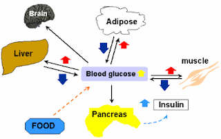
glucose metabolism and homeostasisIn our bodies there are clocks in addition to the Master clock located in the suprachiasmatic nucleus. In computers, there are multiple clocks too, and they are tightly coordinated. For example, Integrated circuits like AV 9155 generate multiple clock frequencies for different portions of a computer (e.g. bus clock, CPU clock, keyboard clock etc.). All these clock frequencies are well regulated, since ICs like AV9155 use 2 quartz crystals (14.318 MHz) which generates of all these frequencies (they have inbuilt circuitry for dividing/multiplying these frequencies to create other necessary frequencies).

Our bodies have their own version of these ‘crystal oscillators’, the BMAL1/CLOCK heterodimer. Since genes are present in all cells (leaving aside germ cells for a while, since they are haploid, and chiasma formation gives rise to gene rearrangement), theoretically all cells also has the machinery for BMAL/CLOCK generation. Thus in the periphery, where these genes are expressed, circadian oscillating mechanisms are automatically incorporated.

The role of peripheral circadian clocks is still uncertain. But it is known that the peripheral clocks regulate cell division, estrous cycles and glucose and lipid homeostasis. Lamia et al knocked out the BMAL1 gene in mice liver and observed that the liver was no longer able to pour sufficient glucose into the blood circulation for cellular activity, resulting in hypoglycemia. Normally, the liver produces glucose from lipids and amino acids in a process called neoglucogenesis; and from glycogen, a glucose polymer, by glycogenolysis, in the fasting phase, to make up for the dwindling blood glucose level. In liver specific BMAL1 deletion, this did not happen and the animal suffered from hypoglycemia, indicating the important role of the liver peripheral clock.

These peripheral clocks certainly need to be regulated too in order to achieve physiological harmony. The master clock in the suprachiasmatic nucleus might regulate these peripheral clocks by hormones and hemodynamic cues.

Gatfield et al used two groups of mice and inactivated BMAL1 in all their cells in one group (BMAL1-/-); and only in liver cells in the other group (L-BMAL1-/-) [the 2 minus signs indicate homozygous, or in both alleles, deletion/inactivation]. The mice in which all BMAL1 were deleted did not show any problem which glucose homeostasis, whereas those with only liver specific BMAL1 deletion had problem maintaining normal sugar level in the inactivity (fasting) phase. Thus the role of liver clock is undeniable. The hepatic oscillator synchronises on feeding cues, since feeding is related to circadian metabolism. In the L-BMAL1 knockout mice, both neoglucogenesis and glycogenolysis operated adequately, but the machinery for the pouring of glucose into the circulation, the final step that is carried out by glucose transporter 2 (GLUT2) is suboptimal. GLUT2 expression in L-BMAL1-/- rats is inadequate.

In BMAL1-/- mice, the master clock in the SCN was inactive along with all other peripheral clocks. This presumably abolished the circadian feeding responses and thus glucose homeostasis was minimally affected. It is as if both the SCN (master) and liver (slave) clocks gone wrong and they were fully asynchronous. But in the L-BMAL1 knockout mice, the SCN was OK and it expected the desired blood glucose level in the habitual feeding time, but the liver lacked GLUT2 to supply the required glucose in the bloodstream. UNITED WE STAND, we better synch!

Last modified: never
References:
Physiological significance of a peripheral tissue circadian clock. Katja A. Lamia, Kai-Florian Storch, and Charles J. Weitz  doi:10.1073/pnas.0806717105

BMAL1 and CLOCK, Two Essential Components of the Circadian Clock, Are Involved in
Glucose Homeostasis. R. Daniel Rudic , Peter McNamara , Anne-Maria Curtis, Raymond C. Boston, Satchidananda Panda, John B. Hogenesch, Garret A. FitzGerald doi:10.1371/journal.pbio.0020377

ResearchBlogging.orgD. Gatfield, U. Schibler (2008). Circadian glucose homeostasis requires compensatory interference between brain and liver clocks Proceedings of the National Academy of Sciences, 105 (39), 14753-14754 DOI: 10.1073/pnas.0807861105

November 25, 2008

Molecular Basis of Genetic Switch In The Circadian Clock

circadian clock showing PER, CRY proteins, Bmal and clock
It is said that the early bird gets the worm. So what is it that makes them rise early? Scientists have questioned it for long. It was in 1995, David Welsh, then a graduate student, discovered that individual cells dissected out from the 'suprachiasmatic nucleus' of rats' hypothalamus showed spontaneous oscillations. And this set the ball rolling!

All organisms from simple unicellular to humans have their own clock mechanisms. We for example, have not one but many oscillators. The master clock that oversees all the other clocks is located in a part of the brain called hypothalamus, the suprachiasmatic Nucleus or SCN for short. The clock circuit is based on transcription and translation of a genetic switch that resides in the SCN. In the nucleus, a gene, called the Per1 gene,  produces a protein called PER (for period). Like other proteins, its production is regulated by a promoter sequence of DNA, which is known as E-box. A heterodimer (dimer because it consists of two molecules; hetero because the molecular weights/size is different) consisting of proteins BMAL1 (also known as MOP 3) and CLOCK sit atop the E-box sequence. Together they regulate the Per1 gene (other clock genes like AVP or arginine-vasopressin genes are also regulated)  resulting in the production of PER1 protein. So, in a way the E-box may be considered as the genetic switch and the heterodimer of BMAL1 and CLOCK the regulator.

Lets suppose that Per1 gene is producing PER1 protein. So, the concentration of this protein in the cytoplasm will rise. This PER1 protein will now combine with other clock proteins namely, PER2 protein, CRY 1 and 2 proteins (CRY for cryptochrome) in the cytoplasm; and will finally reach the nucleus. In the nucleus, they inhibit the Bmal1 and Clock heterodimer transcription factor, which will lead to a drop in PER production. Thus, the positive feedback of BMAL1 and CLOCK on Per1 gene; and negative feedback of PER and CRY protein on the BMAL1 and CLOCK heterodimer keep the clock running. See the adjoining figure. Other proteins like TIM (timeless) and CK1e (casein kinase 1 epsilon; it degrades PER proteins) may also play some role. New research however suggests that CRY proteins, particularly CRY1 protein is a stronger repressor of the said heterodimer.

Research by Leloup et al showed that the mRNA of Bmal1 was in antiphase with that of Per and Cry. This was expected, because they are negatively correlated. Else both the proteins would peak at the same time and the periodicity would be lost. They also observed that the phase of the spontaneous circadian rhythm did not lock. This is because, circadian rhythm is very flexible. In humans, the cycle repeats about every 24.2 hours. The circadian clock is reset by light and our circadian apparatus is exquisitively sensitive to lights falling on the retina. The retina sends this light (for synchronization) to the SCN via the retino-hypothalamic tract. This synchronization or entrainment can now 'phase lock' the circadian rhythm.

Clinical implication of circadian (circa=about; dian=day) rhythm is enormous. Our sleep-wake cycle, growth hormone and cortisol secretion are only a few example. A person in whom the circadian period is short will rise early (early bird?) and a 'night owl' will have his/her circadian period short. Curiously, our sleepiness, tendency to sleep and occurrence of REM sleep peaks (resulting from endogenous circadian rhythm) when we are about to rise; and our endogenous clock reaches its peak about 1-3 hrs before our habitual bedtime. They say that it is a natural homeostatic mechanism, so that we fell less sleepy as daytime passes on and etc. But I not convinced.

But one thing I am sure to abide by is this that I won't deprive my SCN its daily dose of sunlight. I will also not expose myself to undue light (from computer monitor etc) at night and go to bed at a reasonably fixed time. Fiddling with these may result in insomnia or excessive somnolence as in night shift workers and in jet lag (due to latitude/time-zone changes).


Last modified: never
Reference: BMC Molecular Biology 2008, 9:41 doi:10.1186/1471-2199-9-ResearchBlogging.orgJ.-C. Leloup (2003). Toward a detailed computational model for the mammalian circadian clock Proceedings of the National Academy of Sciences, 100 (12), 7051-7056 DOI: 10.1073/pnas.1132112100

November 20, 2008

Hearing Involves Sound Physics

Ear showing ossicles, round window and oval window
The way we hear sound is complex. The different attributes of sound (namely, intensity, frequency, the direction from which it is coming etc.) are faithfully perceived in the auditory cortex. The whole procedure may seem rather straightforward, but it is far more complicated than what looks so deceptively simple.

The sound waves (say from an orchestra) impinge on our eardrums. Sound waves are mechanical waves consisting of condensation and rarefaction, things we learned in our school days. These waves then vibrate our eardrums (Tympanic Membrane; TM). The TM is critically damped, meaning any vibration that is set in will stop almost instantaneously. The vibrating TM then transfers its mechanical energy to the oval window, in the membranous labyrinth of the cochlea, via an ossicular chain consisting of three (3) very small bones.

Basilar membrane, organ of Corti and the scala vestibuli,media and tympani, phalangeal cellsThe membranous labyrinth consists of three adjoining tubes coiled side by side (as shown in the figure). If we were to make a section through it, we would find 3 separate compartments within it: Scala vestibuli, Scala media and Scala tympani. Scala vestibuli is connected to Scala tympani at the apex of the cochlea, a place called helicotrema. While Scala tympani contains a fluid called endolymph (a fluid rich in K+ or potassium ions); the other 2 tubes contain perilymph (a fluid very similar to plasma, rich in Na+ and low in K+). The sheer asymmetry in K+ distribution among the two adjacent fluids (endolymph and perilymph) generate an endocochlear potential of about +80mV, endolymph positive when the perilymph is considered as 0 (zero) volt or ground.

As the oval window vibrates, the fluid in Scala vestibuli (perilymph) also vibrates. Sound was traveling in air before it struck the eardrum, but here, we see that they are now propagating in a fluid medium, which has far more inertia than air. The possible impedance mismatch that would happen is compensated by the eardrum itself and the ossicular chain. The mechanical advantage of the lever system of the ossicular chain, together with the ratio of surface areas of TM and the 'oval window', amplifies the force of sound waves about 22 times, so that the total force at the oval window is 22 times than what the TM experienced originally.

Now, vibrations have set up in the Scala vestibuli form the oval window. These vibrations find their way to the Scala media, as the two tubes are separated by only a very thin membrane (Reissner’s membrane). Hence, fluid in the Scala media (endolymph) vibrates whenever the oval window is vibrating. This Youtube video beautifully illustrates it.


The vibrating endolymph sets up a wavy motion in the basilar membrane (BM). The ‘real analysis’ of sound waves starts here! The BM performs real time spectral analysis of sounds it is presented with (analysis of frequencies below 200Hz is skipped though). We normally hear in the frequency range of 20Hz to 20 kHz.

The hair cells in the organ of Corti, our hearing apparatus, are arranged in such a manner along the BM that those near the base of the cochlea will respond to high frequencies; while as we go up to the apex of the cochlea, the BM reacts best at low frequencies. In other words, each part of the BM has its own unique maxima, the frequency at which the BM responds most well. Below 200Hz, there is no such place encoding.

traveling wave in the basilar membrane
Our ears follow another principle. The ‘traveling wave’ spreads quicker near the base of the cochlea and its speed diminishes fast as it goes up. This ensures that a longer stretch is available for the higher frequencies; else the higher frequency part would have been bunched together, creating a loss in the HF range. This non linearity in traveling wave propagation is thus needed.

Generator Potential
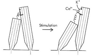
Tip links of hair cells of the earThe genesis of generator potentials in the hair cells is also interesting. Imagine that a group of persons of varying height are standing on a carpet. A thread is attached from the top button (of his shirt) of the smaller person to the top button of his taller counterpart. If the carpet is now tilted by pulling it up from the short person’s end, the thread will now be stretched snapping the taller person’s button. The hair cells also have threads (Tip Links) extending from shorter to their taller cousins. A traveling wave will cause pulling of tip links, resulting in the opening of a mechanically sensitive cation channel. Since potassium is the predominant cation in the endolymph, K+ will then enter the taller hair cells. Look at the electrical gradient around the hair cell; it is -140mV with respect to the endolymph (it is about negative 60 mV wrt the perilymph). So, cations, specially K+ rushes in  creating depolarization. A cascade of events like opening of voltage-gated Ca++ ion channels at the base, consequent fusion and exocytosis of vesicles discharging neurotransmitters, probably glutamate to the afferent cochlear nerve endings surrounding the hair cells, creating an action potential in the nerve.

Frequency Discrimination
When we listen to music, our ears pick up the frequencies in a number of ways. Firstly, the place (maxima) on the BM where maximum excitation takes place is actually a function of frequency. As a matter of fact, there is a frequency map along the BM. Secondly, at frequencies below 3 kHz, the nerves fire in synchrony with the incident sound waves. This is called the ‘volley principle’. The ‘phase locking’ of the two frequencies that occurs below 3 kHz, is highly analogous to the ‘phase locked loops’ in electronic circuits (NE565). Thus ‘volley principle’ allow us to discriminate frequencies. Actually, volley effect is more important in ‘loudness’ assessment. ‘Pitch’ (the subjective/psychological dimension related to frequency) are also moderated by factors such as loudness and the duration of sound. At low frequency (below 500 Hz) pitch seems lower and at higher frequencies (above 4 kHz) pitch seems higher, as the loudness increase, when the frequency is kept constant. Again, when the duration of sound increase from 0.01 second to 0.1 second, the pitch will rise too, for a particular frequency. Sound of less than 0.01 sec duration does not evoke appreciation of any pitch by us.

Loudness Discrimination
Loudness is the perceived intensity of sound, a subjective psychological dimension. The interpreted sound sensation is proportional to the cube root of the actual sound intensity. As such, the ear works at the top of its limit, at a point analogous to Hopf bifurcation, beyond which instability in oscillations occur.
As the sounds become louder, the amplitude of BM movement is more, resulting in more excitation of hair cells. Secondly, with greater loudness, the hair cells around the ‘maxima’ fire too. This causes spatial summation. Thirdly, the outer hair cells are stimulated at loud sounds. The brain will automatically infer loudness levels when cells corresponding to the outer hair cells fire.

Locating Sounds
Then there is location of the direction of sound. We can locate whether the drums are on the left and the lead guitar is to the right. This is achieved by calculating which ear gets the sound first (time lag) and/ or which ear gets it louder (intensity). The time lag method works below 3 kHz; while intensity method works at higher frequencies.
Front/ back discrimination is done by the pinna (auricle) of our ears due to their particular shape.

Auditory nerve
It is interesting that each auditory nerve fiber has its own characteristic frequency, the frequency at which it responds most well. However, it is true only at low intensity. At higher intensities, this specificity is lost and they then respond to a wider spectrum of frequencies. The auditory nerve produces a flurry of action potentials, the frequency of which depends on the intensity of the sound stimuli, it is exposed to. This is very much similar to ‘voltage to frequency converter’ ICs (LM331 is one such VFC IC), where a change in voltage at the input of VFC will cause a change in frequency at the output.

The auditory nerve then goes to the: cochlear nucleus in the medulla to Superior olivary nucleus to Inferior colliculus (via lateral lemniscus) to Medial Geniculate Body (in the Thalamus) to end in Auditory cortex. Some fibers cross to the other side early in their course while others cross at other levels. Fibers from primary auditory area sends association fibers to different parts of the brain for language processing and other tasks.
The nerve synapses with higher order neurons in the above places, which then relay to the nerves upstream. Everywhere in its course, including the nuclei, there are clearcut ‘tonotopic maps’, representing definitive frequency layouts. There are also extensive crossing of nerve fibers to the opposite side. Before the fibers reach the auditory cortex, they connect to many reflex pathways vital to life. For example, they send branches to the reticular activating system which keeps us awake during the noisy hours.

The auditory cortex (Brodmann’s area 41) is the portion of the cerebral cortex in the superior temporal gyrus. Its anterior part is mainly concerned with low frequency and the posterior part tackles the higher frequencies. Thus area 41 also has its own tonotopic map. Secondary auditory cortex (auditory association area) is vital for the interpretation of sounds. A person, in whom Wernicke’s area (part of auditory association area) is damaged, will hear normally but will fail to understand its meaning, leading to aphasia.

The sheer complexity of the auditory circuitry is really mind-boggling: the logarithms (we hear in a log scale, not a linear one), cube functions, phase locking are only some of them. The range of sound intensity (from whisper to the roar of a jet plane) we hear is about 1 trillion fold; but surprisingly, the auditory nerve fibers have a much less dynamic range. Yet we hear the full range. It’s really amazing.

ResearchBlogging.org
P. Martin (2001). Compressive nonlinearity in the hair bundle's active response to mechanical stimulation Proceedings of the National Academy of Sciences, 98 (25), 14386-14391 DOI: 10.1073/pnas.251530498

Last modified: Mar 10, 2014
Reference: Textbook of Medical Physiology, 17e, Guyton and Hall

November 08, 2008

Do We Really Forget? Fathoming The Esoteric Realms of Memory

Smells like teen spirit”, but it could be true that we never really loose any memory in our lifetime. Our memories are stored in the synapses (junction, more specifically, gaps between adjoining neurons) as a function of synaptic strength, in the nerve cells like dendrites as proteins, and some other processes which mostly encompasses a chemical interaction. I am excluding memories such as T cell or B cell memories here; memory, here, will refer to neural ones that occur in the CNS.

We know that in dementias such as global multi infarct dementia, Alzheimer’s disease; there are diffuse losses of neurons and losses of cholinergic neurons in particular, respectively. In surgical cases of epilepsy or brain tumor, there are losses of neurons too. In these cases, memory loss may be irrecoverable, though cases are on record which points to shifting of those memories into some other safe havens. But what about the rest of the population? Does an established long term memory vanish completely?

Let’s consider some facts. The numbers of synapses and their strengths are finite, though both can change in response to stimuli. Even the number of neuron themselves can increase, contrary to the belief held earlier. Neuronal stem cell pool has been identified in the brain. Memories stored in the brain are finite too. Memories are inherently dynamic in nature. Even long term memory stored in the neocortex (medial temporal lobe, on the other hand, stores memories as a buffer, like a D RAM chip, a temporary storage) can change location, as much as transferring itself to the other hemisphere (intercortical transfer), when needed; via the optic chiasm and corpus callosum. So, we see that the number of synapses, though finite, can rise to the demand of an enhanced input from sensory cues which are finite too, leading to memories that can jump across their own allocated territories. A finite brain capacity (say C) can certainly contain a finite memory (say M) as long as C is greater than/ equal to M. Certain computer softwares even trespass this limit; a zip file of 2 MB may deliver 3MB of contents on unzipping! Who knows if the brain isn't using this for the past thousand years.

Synapses, simplistically, may be thought of in binary terms: 1, when it is on; 0, when it is off. Both 1 and 0 is a bit in Boolean terms. We leave aside the synaptic strength part here for the sake of simplicity. In addition, memories may shuttle between synapses in such a way so that it is present in the brain, but not represented by any synapse. I will explain. We all have seen those jugglers juggling those colorful balls too many at a time using only their two hands. A similar thing like dipole dynamics may occur in the brain. Added to this is quantum superposition, which allows the situation of BOTH 1 and 0 state at the same time at the synapse. That the brain can be in a quantum state at the core body temperature and the brain can effectively avoid ‘decoherence’ in the background thermal noise has been discussed by Roger Penrose and Stuart Hameroff. We also know that memories aren’t kept as such, but they are fragmented into individual elements, which are mostly matched to existing elements and are associated. This is economic as it saves space, and useful for indexing and contextual retrieval.

a device for administering deep brain stimulationThus it seems that we ought to have immense memory storage. Haven’t we encountered long forgotten memories in our dreams? Electrical stimulations in some parts of the hippocampus (deep brain stimulation or DBS, figure shown) during routine surgical procedures have given rise to ‘deja vu’ phenomena. The patients remembered things considered long forgotten. We may not be aware of the vast database of memories and are liable to infer that we have “killed ‘em all”, but in reality this may not be the case as Norio Ota et al clearly points it out in their paper. It smells like another chapter from your favorite science fiction novel, but it could be true.

Last modified: never; N.B.There is a substantial amount of speculation in this paper. Please exercise your own judgment and enlighten me about any possible error.
Reference: hyper-links, unless specifically mentioned.

Scientists Simulate Learning In Amoeba Using Memristor

It is surprising how small insects get energy from a wide range of food (not merely petrol or diesel), crawl, fly, reproduce and do so many maneuvers. Now it has been seen that amoeba, a unicellular organism, can learn and memorize too. We are far from creating devices of such versatility, let alone making them as compact as they are.

Amoebae can move, and they do this by changing the physical state they are made of: sol-gel state. The interior of amoebae contains endoplasm, which is in sol state; while the surrounding ectoplasm remains in gel state. The ectoplasm, being in gel state, is more viscous than the inside. When the organism moves, its contractile elements made of actin myofilaments contract, pulling the inside of the amoeba. This causes tension in the endoplasm, creating a change in the sol-gel state. If you squeezed a sponge ball that had been dipped in water, you would notice that water would spurt out from the pores of the sponge. Likewise, the increased tension inside, will create channels through the more viscous ectoplasm, courtesy some parts of ectoplasm (gel state) giving away (to sol state).

We know that reptiles hibernate in winter, when the humidity and temperature is low (we too are no exception 😊). Amoebae too, slow their locomotion in response to these conditions. There are inherent oscillations within the amoeba (alternate sol gel transformation, changes in ionic flux etc) which are continuously adjusted with external signals like temperature and humidity. We, complex multicellular organisms, too have our own master oscillator (circadian clock) in the suprachiasmatic nucleus, which also continuously adjusts by lights falling on the retina.

Yoshiki Kuramoto of Kyoto University and colleagues subjected Physarum polycephalum, an amoeba, to three regularly-spaced dips in temperature and humidity, and found that its locomotive activity decreased. Thereafter, they noticed that a single dip was sufficient to elicit this response. It seems they adjusted their oscillations to the external cue and developed a conditioning later. The study implied that the amoeba anticipated that other such dips might be forthcoming, from the memory it learned. Such response did not occur when the temperature and humidity changes were irregular.

Memory in this case occurs due to the persistence of the channels etched by the organism in the ectoplasm. But this ‘memory’ did not persist for long, if we continued giving them a single dip instead of a regular triplet. This plasticity (change due to reorganization as a function of a stimulus) in amoeba has now been simulated with the aid of electronics by Massimiliano Di Ventra et al.

They used a capacitor, a resistor, an inductor in series and connected a ‘memristor’ in parallel with the capacitor. Memristors (for memory resistors)array of memristors are devices which consist of two layers of titanium dioxide (often present in medicine coatings and chewing gums). When current is applied to one layer, the resistance of the other changes. Leon Chua, of the University of California at Berkeley, predicted it long time ago; and now R. Stanley Williams and colleagues at Hewlett Packard have developed it. It can store memory like DRAM, but unlike DRAM it doesn’t forget when a current is no longer flowing. They hold promise as energy efficient chip for computers and we can also expect faster ‘booting’ of computers, since memory will already have been stored there. The adjoining figure shows ‘memristors’ in a row, as seen by atomic force microscopy (AFM).

Now when a current, fluctuating (AC) in a non periodical manner or a stable DC, was made to pass through the circuit, the memristor went to a low resistance state, virtually short circuiting and dampening the oscillation. However, with a regularly fluctuating current, whose frequency matched the resonant frequency of the circuit, the memristor went into a high resistance state, strengthening the oscillation. What connects electronics to amoeba is the memory that both the circuit retain. The memory of memristor, called memristance, is due to atomic rearrangement in the device. The high resistance state lingers for quite some time, so that next time one single pulse was necessary to put it into oscillation. This phenomenon is quite akin to the protozoal response.

It seems that those days are certainly not far when we will just need to jack-up a USB device in our head to boost up our memory.

Last modified: Nov 13, 2008
Reference: http://arxiv.org/abs/0810.4179?context=q-bio
ResearchBlogging.org Tetsu Saigusa, Yoshiki Kuramoto (2008). Amoebae Anticipate Periodic Events Physical Review Letters, 100 (1) DOI: 10.1103/PhysRevLett.100.018101