Showing posts with label biochemistry. Show all posts

August 16, 2009

Of Twinkling Nanostars and the Possible Application of Stroboscopes in Biological Imaging

Imagine a strong crowd, as you see in a Manchester United versus Liverpool football match and you wished to concentrate on a particular person. How would you do it? Make him wear a fluorescent shirt and dye his hair (don’t do it in the middle of the crowd, I can’t guarantee your safety).

Purdue University researchers have been successful in focusing at the cell of interest among a background of equally noisy and boisterous biomolecules and other metabolically active cells. Currently, researchers use immunological techniques to create an antibody to a molecule and then visualize the ‘molecule of interest’ by tagging the antibody to a radioisotope or a fluorescent dye; and flow cytometry can sort out different types of cells.

The Purdue University team used gold coated nanoparticles with an iron oxide core that was impregnated in the cell they wished to see. They then subjected the specimen to a periodically changing magnetic field. The superparamagnetic cores (superparamagnetic nanoparticles have no net magnetization, but an external magnetic field can magnetize them) responded by rotating as the magnetic field rotated around them. The rotation could be seen in the ‘near infra-red’ light spectrum, as the incident light bounced off (scattered) the specially designed arms of the gold nanostar as it revolved. The rate (rpm) of this gyromagnetic (gyros means to rotate) twinkling could be externally controlled by varying the rate of the externally applied field. You now could identify the cell by its characteristic ‘twinkling’ (lighthouse type) effect.

I am tempted to go beyond what’s been achieved so far. Here I go. I guess you are all familiar what happens to the rotating ceiling fan blades when you turn on a fluorescent lamp. Don’t you see a momentary snapshot of the three blades (some have 4)? That’s what where stroboscope comes in. It consists of a Xenon lamp (ordinary fluorescent lamps could do, but incandescent lamps won’t work as the glowing filament takes time to extinguish) flashing at a controllable rate. The electronic circuitry may be had here.

Suppose that the fan is revolving at 1200 RPM and it is not changing. Set your stroboscope to flash at this rate. You’ll ‘see’ that the fan blades are absolutely not moving, which is certainly not true! But be there any mechanical defect in the fan, it will stand out as the centrifugal force widens it (provided that the fault is more or less tangential to the axis of rotation). Here also we are looking at our object of interest, aren’t we?

Now lets look what implication it might have in biological imaging. We now know that Molecular machine, ATP synthase motorthe gamma subunit of mitochondrial F type ATP Synthase ‘actually’ rotates when it is synthesizing ATP (reverse rotation occurs when ATP is hydrolyzed). There are other locomotive units within the cell as well. They comprise of actin and myosin based molecular motors. Could we study them using an externally adjustable stroboscope? The optical (electromagnetic) signals so obtained may then be similarly broken down into simpler trigonometric (sine and cosine) functions by Fourier analysis (Fourier transform) as was done in the ‘twinkling nanostars’ experiment. At least, we expect to get rid of some 'noise' and some good still photos. But if we wanted better resolution and used higher frequency (electromagnetic) for it, some extraneous error will be introduced. It's a trade-off!

ResearchBlogging.orgLast modified: never
Reference: hyper-links, unless specifically mentioned

Principles of Biochemistry, Lehninger, 4th ed
http://en.wikipedia.org/wiki/ATP_synthase
Wei, Q., Song, H., Leonov, A., Hale, J., Oh, D., Ong, Q., Ritchie, K., & Wei, A. (2009). Gyromagnetic Imaging: Dynamic Optical Contrast Using Gold Nanostars with Magnetic Cores Journal of the American Chemical Society, 131 (28), 9728-9734 DOI: 10.1021/ja901562j
April 05, 2009

Capturing Thought, in Real Time

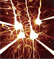
diagram depicting fluorescent optical activity of neurons Wouldn't it be nice if we mapped how the thought processes traveled across our brain, in real time? That's exactly what Mazahir Hasan et al of Max Planck Institute for Medical Research in Heidelberg, have enabled us to view, when an action potential (AP) is underway in the central nervous system (CNS). The researchers introduced fluorescent calcium indicator proteins (FCIP) into the brain cells of mice by means of viral gene vectors. Each time an AP was underway, a lot of ionic phenomena happened. For example, the fast Sodium channels (Na+) opened (letting positive charges to the interior of the cell) leading to depolarization, Potassium (K+) channels opened (to bring back the resting membrane potential to normal, since K+ egress out of the cells) and so on.

Next , the impulse is transmitted to the post-synaptic neuron through the agency of neurotransmitters. But, for this 'coupling' between the presynaptic and postsynaptic neurons to occur; Calcium ion (Ca++) levels in the synaptic knobs of the presynaptic neurons must rise for effective degranulation of the presynaptic vesicles. And that's precisely these researchers were banking upon.

Just before the degranulation of synaptic vesicles begins; calcium ion concentration surges. Such short calcium currents peak within milliseconds, making them the appropriate ions for studying fast neuronal activity. Previously scientists had measured such currents by using microelectrodes implanted within the brain; but this method was quite unsuitable in studying moving animals or for a longer time period. So, they went on to produce stable transgenic mouse lines responding to functional calcium indicators; (including 'inverse pericam' and 'camgaroo-2') using viral vectors. These transgenic mouse lines were under TET inducible promoter (tetracycline, a broad-spectrum antibiotic) control. The TET system offered the advantage of targeting combination of different neuronal cell assemblies. The other side of the Ptetbi (bidirectional promoter tetracycline) promoter was attached to the firefly luciferase gene. They were also sensitive to doxicline (another antibiotic belonging to the same category as tetracycline) in terms of regulation of luciferase, as well.

They then used a heteromeric sensor protein called D3cpv, which was made to produce in the nerve cells of the transgenic mice. Two subunits of this protein reacted to the binding of calcium ions in a way that when the yellow-fluorescent protein (YFP) lit up and the cyan-fluorescent protein (CFP) intensity diminished. When calcium was bound to the D3cpv complex; CFP (cyan fluorescent protein) and YFP (yellow fluorescent protein) came closer together bringing about FRET, in such a way that there was a visible color change, 'visually' or optically indicating the progression of action potential in real time. CFP and YFP are spectral variants of GFP linked together by a Ca++ sensitive linker.

They used 'two-photon imaging microscopy' to study this phenomenon. They excited thinned out rat skulls using two-photons simultaneously using 'mode-locked' Titanium-sapphire laser. They then amplified the signal using photomultipliers and analyzed them.

The resolution of the experiment was limited to less than 1 Hz (frequency of action potentials). They conferred that human thought processes might be mapped in much the same 'opto-physiologic way', in contrast to the usual electrophysiologic approach. Not only does the experiment throw light on the thought processes in real-time, but also, it is expected that it will be useful in the pathophysiology and treatment of Alzheimer's disease, Parkinson's disease and Huntington's chorea.

FCIP-positive cells were found in the hippocampal CA1 and CA3 regions, mossy fiber areas of the dentate gyrus, neocortical pyramidal cells and olfactory receptor neurons, they remarked. They studied cortical pyramidal cell, olfactory and optical responses in the mice in their experiment.

ResearchBlogging.orgHasan, M., Friedrich, R., Euler, T., Larkum, M., Giese, G., Both, M., Duebel, J., Waters, J., Bujard, H., Griesbeck, O., Tsien, R., Nagai, T., Miyawaki, A., & Denk, W. (2004). Functional Fluorescent Ca2+ Indicator Proteins in Transgenic Mice under TET Control PLoS Biology, 2 (6) DOI: 10.1371/journal.pbio.0020163
Last modified: never
Reference: Damian J Wallace, Stephan Meyer zum Alten Borgloh, Simone Astori, Ying Yang, Melanie Bausen, Sebastian Kügler, Amy E Palmer, Roger Y Tsien, Rolf Sprengel, Jason N D Kerr, Winfried Denk & Mazahir T Hasan. doi:10.1038/nmeth.1242

March 26, 2008

Toward An Objective Correlate Of Pain

visual analog scaleWhile taking a hot water bag to find relief for a severe spondylosis pain, I wondered why pain could not be expressed in a way different from the generally used Visual analog scale. The patient is asked to look at a chart (shown in the figure) and told to rate his sensation, that tallies most well with his pain. If that hot bag were to be applied to a person not having any pain, he would jump almost instantly. The fact that I tolerated it so well (and benefited from it), only shows its countering (counter irritant property) property, which should be somewhat proportional to the severity of one's pain. The relationship of a counter-irritant to pain severity, whether linear, logarithmic or exponential, needs to be established and quantified.

A patient's own account of pain may be subjectively modified according to the personality of the patient and many other factors. As such, this type of quantification is liable to be erroneous. Pain should better be measured in an objective manner, free of bias. In this instance cited above, one could use the formula: Q(heat)=m(mass) x s(specific heat of the substance) x t(temperature), to know the amount of heat energy transferred to the patient. I am assuming that the pain relieving techniques will, kind of, obey Newton's Third law. Amount (intensity) of counterirritant that just suffices pain relief will be equal to the degree of pain. But it is not actually so, as we will see later.

By noting the difference in local temperatures before and after the procedure, one could get
"t".
Mass or "m" could be measured by estimating the volume of the body tissue that was actually heated by infrared mapping, for example; and the expected density of that area. Specific heat for the tissue in question could be easily known and standardized using some cross-sectional studies. A suitable nomogram may later be drawn by plotting values obtained from such observations, for quick estimates. It seems logical that pain so measured, will have its units in British Thermal Units (btu), calories or their work (mechanical) equivalents like ergs, Joules, foot-pounds etc..

In pain therapies using mechanical energies (Ultrasound), electromagnetic devices (laser, short wave diathermy, high frequency photons such as X rays) , a similar formula may be used to obtain the pain equivalent. For example, in laser or short wave therapy, we may design a device that will measure the amount of energy in Watt.seconds/Joules the given area of tissue is supposed to absorb, over a given period of time. The chemical analgesics (pharmaceuticals e.g. Non Steroidal Anti Inflammatory Drugs or steroids; counter irritants such as capsaicin) may be quantified using Scoville scale or by evaluation on the degree of relief from algesia.

Calculating pain may be quite painful in itself. Pain sensation does not tally linearly with noxious stimulus. Rather, a logarithmic relationship was proposed in the Weber Fechner law, which held that the magnitude of pain (or a sensation) felt, was proportional to the log of the intensity of a stimulus. In other words, to feel twice as much pain, you needed to hurt 10 times! To complicate matters further, our present knowledge suggests that the magnitude of a sensation is related by a power factor to the intensity of that sensation. R=KS^A; where R is the sensation felt, S the intensity of stimulus, K and A are constants for that particular tissue. The brighter side is, we get a preformulated relationship for pain calculation.

Pain (musculo skeletal/ visceral, exogenous/endogenous) is generally of two types: fast pain and slow pain. Fast pains such as sharp pain of pricks, stabs are usually carried by Ad (A delta) nerve fibres, while slow aching pains are carried by type C nerve fibers. Ad fibres can carry impulses rapidly as these fibers are myelinated and are of large caliber. C fibers, on the other hand, are unmyelinated and narrow. A-delta fibres release glutamate and C fibers secrete substance-P. A way to measure these chemicals could be a step closer to quantifying pain.

The signals from these fibers travel to the thalamus, a part of the lower brain, on their way to the cerebrum for the localization of pain. Measuring the metabolic activity in thalamus, arising out of increased neural discharges there, by fMRI or PET scan may also shed some light on the intensity of pain stimuli (the stimulus at this level is unmodified by the higher brain) that reaches thalamus. We can also measure the blood levels of endogenous opioids (enkephalins, endorphins) which are secreted in body's response to the pain and adrenaline, secreted in response to increased sympathetic discharge, which is an usual accompaniment of pain. Their blood/plasma levels may correspond with pain severity. Other pain markers like bradykinin, histamine, potassium ions and proteolytic enzymes could be probed too.

True, that the patient may or may not feel as much pain as has been measured this way, because pain perception may not be proportional to the physical/chemical parameters thus described and it is not uniform in all subjects. The brain sees pain in its own mathematical terms, and everyones' brain is different in this regard. A soldier may overlook his gushing wounds, whereas pampered girls of rich persons may feel a lot of pain from an apparently trivial injury. But, quantification of pain in this way (by measuring the physical/chemical yardsticks) may correctly establish the severity of pain in silent myocardial infarction of neuropathy (pain sensation is dulled here due to neural malfunction), decubitus ulcers, trophic ulcers, malingering and in similar situations. In this way we may be able to find a better and objective correlate of pain in clinical practice and develop more efficient analgesics.

P.S. In a recent development, some objective physiological correlates of pain has been tracked. These include measurements from the nonlinear composite of heart rate, heart rate variability, amplitude of the photoplethysmogram, skin conductance, fluctuations in skin conductance, and their time derivatives.  Algorithms can then convert the data into a real-time, continuous index on a bedside monitor. This has resulted in the fabrication of a wearable sensing device can be mounted on a finger.

Last modified: Nov 28, 2015
Reference: hyper-links, unless specifically mentioned

March 11, 2008

Duroquinone: A Parallelly Processing Chemical Computer

Duroquinone nanobots constituting chemical brainDr Anirban Bandyopadhyay of the National Institute for Materials Science, Tsukuba, Japan, have developed a tiny chemical nano-brain, that could one day be guided by remote control. These machines could make surgery on human bodies easier and help revolutionize the computing power of future computers.

Scientists have built nanobots (nanoscale robots; nano means a billionth of a metre) previously but these bots could not be controlled by outside means. Dr. Bandyopadhyay has now devised a nanobot, a chemical one and not mechanical or electronic one, that can be controlled from outside.

This promising nanomachine, just 2 billionths of a meter across, consists of a molecule called duroquinone. A single nanomachine comprises of 17 duroquinone molecules; with one molecule at the center and remaining 16 surrounding it. All these molecules are connected by hydrogen bonding. As is shown in the figure, each duroquinone molecule has four spoke like arms jutting out from it, which can be independently rotated to represent four different states. Thus they can be made to represent four different 'logic states', bits: 0,1,2,3. While ordinary computers work on binary logic (0,1), computers using this technology would have four billion possible combinations with this chemical brain.

The molecule at the center, to which the rest are connected, can be controlled by a scanning tunneling microscope (STM). This machine is not only capable of 'manipulating/directing' their (nanobots') orientations, it is also capable of 'reading' the states they are in. They act rather like both a transducer and a receiver. By tweaking the central molecule, one could switch the nanobot's configurations. Comparable switches in electronic circuits include CD4066, a quad bilateral switch, electrical relays, transistors and others. But here, we are controlling a chemical device by using STMs. In future, we may be able to operate the duroquinone machinery by using the conformational properties of proteins, by optical devices like lasers and may be other electromagnetic devices too.

The researchers were inspired by the parallel processing circuitry of the glial cells in the brain.
Said Dr. Bandyopadhayay, "Doctors will inject molecular machines attached to similar control unit, the assembly will go to the target part inside our body through veins, and carry out bloodless surgery. Till now several molecular machines have been built, prior to this work, but there were no machine that could control them."

March 06, 2008

Nano Does a Mega Tango: FETs To Change The Fate Of Diabetics

The role of electronics in medicine or physiology is a very important one. Ranging from CT scan, fMRI, PET scan or electron microscopes and a variety of other medical investigations, medical science depends on electronics and its allied disciplines.

A simple illustration is as follows. Everyone knows that the estimation of blood glucose is the cornerstone in the diagnosis and therapy of diabetes mellitus and other hyperglycemic illnesses such as hyperthyroidism, hyperpituitarism and others. Even hypoglycemic ailments (conditions where the blood glucose levels are less than normal) like hypothyroidism, hypopituitarism or hyperinsulinemia, hypoadrenocorticism need the estimation or quantitation (quantification) of blood glucose, in order to clinch their diagnosis.

Glucose may be estimated in the blood in a variety of methods. One of the easiest methods rely on the simple reaction of glucose with potassium per-manganate. Glucose is a reducing monosachharide; while potassium permanganate is a strong oxidizing agent, so much so that it produces frank fire when added to glycerine not to mention of its emanation of oxygen (O2) when heated. When KMnO4 (potassium permanganate) is added to glucose, it (KMnO4) is reduced to manganese ions (Mn++). The colour changes from purple pink to colorless. This can be seen by the naked eye, giving us an estimate of the amount of total glucose that is present.

Another approach that is adopted nowadays is the enzymatic oxidation of glucose and their quantitation thereof. Here enzyme glucose oxidase (GOD) is used to oxidize glucose to D-gluconic acid and hydrogen peroxide (H2O2). In the presence of H2O2, hydrogen peroxidase (POD) oxidizes phenol, which combines with 4-amino antipyrine to form a red dye (quinone imine). The intensity of the red dye is linearly proportional (upto 500 mg% i.e. 500 mg per dl) to the glucose concentration in the specimen. The intensity of the color that is formed is measured by using a colorimeter; a device that measures the transmittivity of light (through the liquid) by employing LDR (Light Dependent Resistor) or phototransistors or similar devices.

Recently, advances in nanotechnology and nanoscience as a whole, is offering a brand new hope of determining blood glucose within the patients blood itself. That is you don't need to prick the guy. They are using a device called ISFET (Ion Sensitive Field Effect Transistor).FET with its symbol and pinouts It is variant of FET devices (a picture of a MOSFET i.e metal oxide semiconductor FET, is shown on the left). In a ordinary bipolar transistor, we forward bias the emitter-base junction by applying a current and make the collector strongly reverse biased. Thus any input current in the base emitter circuit is amplified several times when the electrons 'rushes' towards the attracting collector. A FET also works in much the same way. The difference is that the flow of charge carriers are controlled by a electric field applied in the Gate (G) Source (S) junction. This electric field is generated by a voltage: VGS. Thus while bipolar junction transistors (BJT) are current controlled, the FET devices are voltage controlled.

Recently, a team of physicists led by Raj Mohanty from Boston University has made a nanoscale glucose sensor using ISFETs and nanowires consisting of silicon. The silicon nanowires were primed with glucose oxidase on their surfaces and these nanowires bridged the source and the drain electrode of the MOS device. The conductivity of the 'wire' would change depending upon the glucose concentration in blood as it is reacted upon by the enzyme present on its surface. A voltage drop will ensue between the source and the drain electrodes, which may be read and interpreted by a voltage comparator. The researchers claimed that these could be put in vivo with virtually no risk. Humans do have the habit of carrying silicon on their bodies and that hardly do them any harm! We may be able to noninvasively monitor the blood glucose of diabetic persons, may even be able to act upon it, by incorporating a feedback circuit that would actuate an insulin pump, in times of hyperglycemia.

November 20, 2007

Of Mighty Mice and Men

picture of a mouse alongside a cordless mouseIn my article, Doping: To dope or not to dope, I mentioned how athletes exploit the pharmaceutical and other abilities of drugs to manipulate things in their favor. Trained athletes learn to use fatty acids as energy source more effectively than untrained men, over time. Burning fatty acids as fuel has the advantage of not producing lactate, metabolites of carbohydrates that is the cause of muscle cramps. At the same time, lactate, being acidic in nature, inhibit vital enzymes necessary for tissue reactions. Trained (and trendy) athletes who are experienced, overfeed themselves with carbohydrates days prior to a sporting event. This produces ample glycogen stores in them which come handy during the event as energy source.

But it seems that the mouse is going to beat them at their own turf! I am not talking about your brand new electronic optical (or bluetooth) mouse, I refer to our own natural rodent. It is well known that scientists have a penchant for using mice as experimental guinea pigs for their research. In this process they have produced "brainbow" in mice, a multicolored novel approach to explore neural networking; given birth to fearless breed of mice-no longer afraid of cats, by ablating their olfactory (smell) neurones and doing away with their smell associated identification of their enemies etc.

Now scientists have inserted a gene into mice that has made them far more stronger, hardier and has more stamina. This research was led by Richard Hanson, professor of biochemistry at Case Western Reserve University at Cleveland, Ohio. He first made a cDNA (complimentary DNA) of the enzyme PEPCK-C (phosphoenol pyruvate carboxykinase). Creating a cDNA may be compared to making a rubber stamp (or the negative of a photograph) which when inserted into the DNA sequence will keep on 'stamping', creating more proteins (enzymes) in the process. PEPCK-C cDNA was then linked to the skeletal actin gene promoter (a promoter may be conceived as a DNA sequence that instructs a gene to transcribe). Thus PEPCK-C was produced along with actin and the "mighty mouse" emerged. The PEPCK-C gene is expressed in the liver, kidney and some other tissues. This enzyme plays vital role in both carbohydrate and lipid metabolism, the transgenic mice that were produced was very efficient in utilizing fatty acids as energy source.

Cause for concern? Well, I can't envisage a mouse brigade in the near future.

November 19, 2007

Chiral Delight

arrangement of rings to exemplify chiral structuresThe word Chiral comes from the Greek word meaning hand ('Cheiro' the great palmist also got his name from the Greek word). Like those molecules described in Chiral Fallacy, many of our biomolecules in physiology remain in either of these two states. If you were to shine a plane-polarized light through a liquid containing this molecule, the molecules would rotate the beam of light either in a right or left direction.

Just a DIY. Take a polarizer (You may get one from a camera shop or you may get one from your mobile phone LCD display!). Now hold one in a fixed position while you rotate the other over it and watch the translucency change. This occurs due to obliteration of the wave functions of light, as the electromagnetic wave gets chopped off by the two consecutive polarizers. If you place a bottle of water between these polarizers, the light will remain where it was. Replace water with dextrose, a sugar, you will find that the beam now rotates to the right!

Substances which rotate plane-polarized light to the right are termed (+) whereas those rotating towards the left are termed (-). The Cahn-Ingold-Prelog system is commonly used to describe chiral molecules as R (rectus) or S (sinister). An equimolar mixture of both will not rotate the beam. Such a mixture is called racemic mixture.

The thyroid gland, for example, secretes the L (levo) isomer of thyroxine. The D (dextro) enantiomer has little activity but has cholesterol lowering activity instead. Chiral drugs like levocetirizine (used in allergy), S-amlodipine (a calcium channel blocker cardiovascular drug used in hypertension), esomeprazole (a drug for peptic ulcer) or levofloxacin (an antibiotic) are some popular examples.

New drug developments will target this phenomenon of optical isomerism and exploit eutomers (active enantiomers are called eutomers and inactive counterparts are distomers) for more specific effect and less adverse reactions.

November 12, 2007

The Chiral Fallacy

diagrammatic representation of chiral moleculesDo you wear your left gloves on your right hand? I know you don't. It is a fact that though the gloves are almost identical they are not interchangeable. One is the mirror image (copy) of the other. So, wearing your left gloves on your right hand is wrong (isn't left always wrong?).

When a molecule docks on another, to bind, it is said that the molecule has an affinity for it. It may (or may not) elicit a response (efficacy), as a result of this combination. But for this molecule in question; be it a hormone, an enzyme or a medicine, to combine with its consort (receptor/substrate), the molecule needs to embark on the substrate first. In a three dimensional world (leaving aside time dimension for a moment), the molecule (ligand/enzyme) has to use at least 3 points (bonds) to anchor. As shown in the figure, the pyramidal looking molecules display mirror image symmetry. Think of the black balls as indicating depth; the other three balls rest on the substrate molecule like a tripod, in a condition called 3-point attachment.

Now imagine that you are holding the black ball and placing the 'planar tripod' on three imaginary holes (of complementary colors), which correspond spatially to the three projections. No problem. You try to repeat the same thing with the other pyramid, you can't get your jigsaw done! This occurs due to the presence of an asymmetric carbon atom.

This is because though the molecules have the same molecular structures their orientations are different. This is known as isomerism, a phenomenon akin to the gloves analogy. Isomerism has immense importance in human physiology, biochemistry and pharmacology.