Showing posts with label circadian rhythm. Show all posts

December 03, 2008

Peripheral Clocks Synch With The Master Zeitgeber

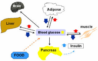
glucose metabolism and homeostasisIn our bodies there are clocks in addition to the Master clock located in the suprachiasmatic nucleus. In computers, there are multiple clocks too, and they are tightly coordinated. For example, Integrated circuits like AV 9155 generate multiple clock frequencies for different portions of a computer (e.g. bus clock, CPU clock, keyboard clock etc.). All these clock frequencies are well regulated, since ICs like AV9155 use 2 quartz crystals (14.318 MHz) which generates of all these frequencies (they have inbuilt circuitry for dividing/multiplying these frequencies to create other necessary frequencies).

Our bodies have their own version of these ‘crystal oscillators’, the BMAL1/CLOCK heterodimer. Since genes are present in all cells (leaving aside germ cells for a while, since they are haploid, and chiasma formation gives rise to gene rearrangement), theoretically all cells also has the machinery for BMAL/CLOCK generation. Thus in the periphery, where these genes are expressed, circadian oscillating mechanisms are automatically incorporated.

The role of peripheral circadian clocks is still uncertain. But it is known that the peripheral clocks regulate cell division, estrous cycles and glucose and lipid homeostasis. Lamia et al knocked out the BMAL1 gene in mice liver and observed that the liver was no longer able to pour sufficient glucose into the blood circulation for cellular activity, resulting in hypoglycemia. Normally, the liver produces glucose from lipids and amino acids in a process called neoglucogenesis; and from glycogen, a glucose polymer, by glycogenolysis, in the fasting phase, to make up for the dwindling blood glucose level. In liver specific BMAL1 deletion, this did not happen and the animal suffered from hypoglycemia, indicating the important role of the liver peripheral clock.

These peripheral clocks certainly need to be regulated too in order to achieve physiological harmony. The master clock in the suprachiasmatic nucleus might regulate these peripheral clocks by hormones and hemodynamic cues.

Gatfield et al used two groups of mice and inactivated BMAL1 in all their cells in one group (BMAL1-/-); and only in liver cells in the other group (L-BMAL1-/-) [the 2 minus signs indicate homozygous, or in both alleles, deletion/inactivation]. The mice in which all BMAL1 were deleted did not show any problem which glucose homeostasis, whereas those with only liver specific BMAL1 deletion had problem maintaining normal sugar level in the inactivity (fasting) phase. Thus the role of liver clock is undeniable. The hepatic oscillator synchronises on feeding cues, since feeding is related to circadian metabolism. In the L-BMAL1 knockout mice, both neoglucogenesis and glycogenolysis operated adequately, but the machinery for the pouring of glucose into the circulation, the final step that is carried out by glucose transporter 2 (GLUT2) is suboptimal. GLUT2 expression in L-BMAL1-/- rats is inadequate.

In BMAL1-/- mice, the master clock in the SCN was inactive along with all other peripheral clocks. This presumably abolished the circadian feeding responses and thus glucose homeostasis was minimally affected. It is as if both the SCN (master) and liver (slave) clocks gone wrong and they were fully asynchronous. But in the L-BMAL1 knockout mice, the SCN was OK and it expected the desired blood glucose level in the habitual feeding time, but the liver lacked GLUT2 to supply the required glucose in the bloodstream. UNITED WE STAND, we better synch!

Last modified: never
References:
Physiological significance of a peripheral tissue circadian clock. Katja A. Lamia, Kai-Florian Storch, and Charles J. Weitz  doi:10.1073/pnas.0806717105

BMAL1 and CLOCK, Two Essential Components of the Circadian Clock, Are Involved in
Glucose Homeostasis. R. Daniel Rudic , Peter McNamara , Anne-Maria Curtis, Raymond C. Boston, Satchidananda Panda, John B. Hogenesch, Garret A. FitzGerald doi:10.1371/journal.pbio.0020377

ResearchBlogging.orgD. Gatfield, U. Schibler (2008). Circadian glucose homeostasis requires compensatory interference between brain and liver clocks Proceedings of the National Academy of Sciences, 105 (39), 14753-14754 DOI: 10.1073/pnas.0807861105

November 25, 2008

Molecular Basis of Genetic Switch In The Circadian Clock

circadian clock showing PER, CRY proteins, Bmal and clock
It is said that the early bird gets the worm. So what is it that makes them rise early? Scientists have questioned it for long. It was in 1995, David Welsh, then a graduate student, discovered that individual cells dissected out from the 'suprachiasmatic nucleus' of rats' hypothalamus showed spontaneous oscillations. And this set the ball rolling!

All organisms from simple unicellular to humans have their own clock mechanisms. We for example, have not one but many oscillators. The master clock that oversees all the other clocks is located in a part of the brain called hypothalamus, the suprachiasmatic Nucleus or SCN for short. The clock circuit is based on transcription and translation of a genetic switch that resides in the SCN. In the nucleus, a gene, called the Per1 gene,  produces a protein called PER (for period). Like other proteins, its production is regulated by a promoter sequence of DNA, which is known as E-box. A heterodimer (dimer because it consists of two molecules; hetero because the molecular weights/size is different) consisting of proteins BMAL1 (also known as MOP 3) and CLOCK sit atop the E-box sequence. Together they regulate the Per1 gene (other clock genes like AVP or arginine-vasopressin genes are also regulated)  resulting in the production of PER1 protein. So, in a way the E-box may be considered as the genetic switch and the heterodimer of BMAL1 and CLOCK the regulator.

Lets suppose that Per1 gene is producing PER1 protein. So, the concentration of this protein in the cytoplasm will rise. This PER1 protein will now combine with other clock proteins namely, PER2 protein, CRY 1 and 2 proteins (CRY for cryptochrome) in the cytoplasm; and will finally reach the nucleus. In the nucleus, they inhibit the Bmal1 and Clock heterodimer transcription factor, which will lead to a drop in PER production. Thus, the positive feedback of BMAL1 and CLOCK on Per1 gene; and negative feedback of PER and CRY protein on the BMAL1 and CLOCK heterodimer keep the clock running. See the adjoining figure. Other proteins like TIM (timeless) and CK1e (casein kinase 1 epsilon; it degrades PER proteins) may also play some role. New research however suggests that CRY proteins, particularly CRY1 protein is a stronger repressor of the said heterodimer.

Research by Leloup et al showed that the mRNA of Bmal1 was in antiphase with that of Per and Cry. This was expected, because they are negatively correlated. Else both the proteins would peak at the same time and the periodicity would be lost. They also observed that the phase of the spontaneous circadian rhythm did not lock. This is because, circadian rhythm is very flexible. In humans, the cycle repeats about every 24.2 hours. The circadian clock is reset by light and our circadian apparatus is exquisitively sensitive to lights falling on the retina. The retina sends this light (for synchronization) to the SCN via the retino-hypothalamic tract. This synchronization or entrainment can now 'phase lock' the circadian rhythm.

Clinical implication of circadian (circa=about; dian=day) rhythm is enormous. Our sleep-wake cycle, growth hormone and cortisol secretion are only a few example. A person in whom the circadian period is short will rise early (early bird?) and a 'night owl' will have his/her circadian period short. Curiously, our sleepiness, tendency to sleep and occurrence of REM sleep peaks (resulting from endogenous circadian rhythm) when we are about to rise; and our endogenous clock reaches its peak about 1-3 hrs before our habitual bedtime. They say that it is a natural homeostatic mechanism, so that we fell less sleepy as daytime passes on and etc. But I not convinced.

But one thing I am sure to abide by is this that I won't deprive my SCN its daily dose of sunlight. I will also not expose myself to undue light (from computer monitor etc) at night and go to bed at a reasonably fixed time. Fiddling with these may result in insomnia or excessive somnolence as in night shift workers and in jet lag (due to latitude/time-zone changes).


Last modified: never
Reference: BMC Molecular Biology 2008, 9:41 doi:10.1186/1471-2199-9-ResearchBlogging.orgJ.-C. Leloup (2003). Toward a detailed computational model for the mammalian circadian clock Proceedings of the National Academy of Sciences, 100 (12), 7051-7056 DOI: 10.1073/pnas.1132112100

November 08, 2008

Scientists Simulate Learning In Amoeba Using Memristor

It is surprising how small insects get energy from a wide range of food (not merely petrol or diesel), crawl, fly, reproduce and do so many maneuvers. Now it has been seen that amoeba, a unicellular organism, can learn and memorize too. We are far from creating devices of such versatility, let alone making them as compact as they are.

Amoebae can move, and they do this by changing the physical state they are made of: sol-gel state. The interior of amoebae contains endoplasm, which is in sol state; while the surrounding ectoplasm remains in gel state. The ectoplasm, being in gel state, is more viscous than the inside. When the organism moves, its contractile elements made of actin myofilaments contract, pulling the inside of the amoeba. This causes tension in the endoplasm, creating a change in the sol-gel state. If you squeezed a sponge ball that had been dipped in water, you would notice that water would spurt out from the pores of the sponge. Likewise, the increased tension inside, will create channels through the more viscous ectoplasm, courtesy some parts of ectoplasm (gel state) giving away (to sol state).

We know that reptiles hibernate in winter, when the humidity and temperature is low (we too are no exception 😊). Amoebae too, slow their locomotion in response to these conditions. There are inherent oscillations within the amoeba (alternate sol gel transformation, changes in ionic flux etc) which are continuously adjusted with external signals like temperature and humidity. We, complex multicellular organisms, too have our own master oscillator (circadian clock) in the suprachiasmatic nucleus, which also continuously adjusts by lights falling on the retina.

Yoshiki Kuramoto of Kyoto University and colleagues subjected Physarum polycephalum, an amoeba, to three regularly-spaced dips in temperature and humidity, and found that its locomotive activity decreased. Thereafter, they noticed that a single dip was sufficient to elicit this response. It seems they adjusted their oscillations to the external cue and developed a conditioning later. The study implied that the amoeba anticipated that other such dips might be forthcoming, from the memory it learned. Such response did not occur when the temperature and humidity changes were irregular.

Memory in this case occurs due to the persistence of the channels etched by the organism in the ectoplasm. But this ‘memory’ did not persist for long, if we continued giving them a single dip instead of a regular triplet. This plasticity (change due to reorganization as a function of a stimulus) in amoeba has now been simulated with the aid of electronics by Massimiliano Di Ventra et al.

They used a capacitor, a resistor, an inductor in series and connected a ‘memristor’ in parallel with the capacitor. Memristors (for memory resistors)array of memristors are devices which consist of two layers of titanium dioxide (often present in medicine coatings and chewing gums). When current is applied to one layer, the resistance of the other changes. Leon Chua, of the University of California at Berkeley, predicted it long time ago; and now R. Stanley Williams and colleagues at Hewlett Packard have developed it. It can store memory like DRAM, but unlike DRAM it doesn’t forget when a current is no longer flowing. They hold promise as energy efficient chip for computers and we can also expect faster ‘booting’ of computers, since memory will already have been stored there. The adjoining figure shows ‘memristors’ in a row, as seen by atomic force microscopy (AFM).

Now when a current, fluctuating (AC) in a non periodical manner or a stable DC, was made to pass through the circuit, the memristor went to a low resistance state, virtually short circuiting and dampening the oscillation. However, with a regularly fluctuating current, whose frequency matched the resonant frequency of the circuit, the memristor went into a high resistance state, strengthening the oscillation. What connects electronics to amoeba is the memory that both the circuit retain. The memory of memristor, called memristance, is due to atomic rearrangement in the device. The high resistance state lingers for quite some time, so that next time one single pulse was necessary to put it into oscillation. This phenomenon is quite akin to the protozoal response.

It seems that those days are certainly not far when we will just need to jack-up a USB device in our head to boost up our memory.

Last modified: Nov 13, 2008
Reference: http://arxiv.org/abs/0810.4179?context=q-bio
ResearchBlogging.org Tetsu Saigusa, Yoshiki Kuramoto (2008). Amoebae Anticipate Periodic Events Physical Review Letters, 100 (1) DOI: 10.1103/PhysRevLett.100.018101

September 16, 2007

Electronic versus Biological Clock: A Comparative Analysis

Electronic clockElectronic clocks rely on oscillators to derive its clock signals. Modern day clocks employ crystals made of quartz, instead of employing CR (Capacitor-Resistor) or LC (Inductor(L)-Capacitor) networks, transistors or 'flip flop' integrated circuits, for the production of clock signals. In a typical electronic device, such as a computer or mobile phones, there is the need for not one but many different clock signals, for its different sub-systems. This is achieved by using decade counters (such as the TTL IC 7490, TTL stands for transistor transistor logic), which divide the clock frequencies by 10, or by using dividing by 12 IC 7492. By using such devices and others, in cascading manners, many different frequencies can be synthesized which may then be fed to the corresponding sub-systems.

In both plant and animal kingdoms, including humans, there are many 'clock generating' cells that functions as biological clocks. For example, there is one such clock in the hypothalamus (a part of our brain), which goes by the name of 'circadian clock'. This clock, also called the master clock, is actually a conglomeration of neurones at suprachiasmatic nucleus (SCN) of the hypothalamus, which produces its clock cycles in more or less the same manner as an electronic oscillator does. Circadian means about (circa) days (dies), in Latin, and as the name suggests, this clock enables important tasks, those have some relevance with the time of the day, to be executed in time. Activities such as release of some hormones (ACTH or adrenocorticotrophic hormone, cortisol etc), regulation of body temperature, sleep-wake cycle, behavior, growth and many other activities are controlled by the circadian clock and hence these activities comprise what we call circadian or diurnal rhythms. Like electronic clocks, they are also fed by reset signals, so that the clock may be synchronized with external stimuli, called 'zeitgebers', German for time givers. During daylight, light falls on the retina, the ganglion cells there are stimulated via their melanopsin photoreceptors, which then send this information to the SCN ('master clock') through the retino-hypothalamic tract. This way the clock remains up to date. This story of relay doesn't end here. The hypothalamus again relays this cue to pass on to the pineal gland, which produces 'melatonin' that plays important role in sleep wake cycle.
There other clocks in the brain too. For example, in the arcuate nucleus of the hypothalamus, a pulsatile clock, controls GnRH (gonadotrophin releasing hormone) secretion. This hormone, in turn, regulates the secretion of gonadotrophins: Leutinizing and Follicle stimulating hormone (LH & FSH), which are instrumental in reproduction. Then there are 'peripheral clocks' in areas such as the heart (cardiac pacemaker), intestinal smooth muscles, liver and other organs. In addition, each individual nucleated cell has its own in-built telomere operated clock, that counts 50 times after which the cell self apoptoses (Hayflick limit). This clock is DNA operated. It is likewise speculated that the circadian clock may also be DNA operated, and DNA transcription/translation feedback mechanism, may be the one that does the counting trick. Barring some exceptions, the ultimate effector is change in ionic fluxes in the neurones.

Whatever may be the mechanism of genesis of the clock pulses, it is to be borne in mind that these numerous clocks have to be coordinated, to be of relevance in physiology. This coordination is done sometimes through neuroendocrine mechanisms and sometimes through other means. Since ionic fluxes are involved, it may be possible that in the brain, where electrical circuits abound, electromagnetic coupling might play some role. Also, if the distance between the transmitting and receiving units is negligible and the chances of 'decoherence' is small, coupling through quantum mechanism by entanglement is also not an untenable option. Therefore we need to be open minded and logical if we were to come to a meaningful and universally accepted theory, before the sands of time run out.