Showing posts with label nuclear physics. Show all posts

September 11, 2010

fMRI, BOLD and the Beautiful

ResearchBlogging.orgWhen we want to examine the brain of a person noninvasively by Computed Tomography (CT) or MRI, we get a ‘snapshot’ of the anatomy (or pathology, if any) of the subject’s brain. We are however clueless as to its functional aspect. fMRI or Functional Magnetic Resonant Imaging allows us to do just that. The difference is not unlike a ‘still picture’ versus a ‘video of a moving train’. PET scans, previously described, also can asses the functional state of the brain.

Whenever we do a task, think, dream, memorize, speak or see things, the brain is not activated as a whole; but only certain portions of it are activated. Activation, here, means increased metabolic activity of neurons in certain areas of the brain. Naturally, these ‘metabolically active’ neurons would demand more energy which would power them. The blood supply to these areas increases as a result of this metabolically driven vasodilation. The arteries then bring in glucose and oxygen with them, with Oxygen being transported in the form of Oxyhemoglobin (oxygenated hemoglobin or HbO2). Neurons on the other hand use up the oxygen contained in the blood, thereby reducing it to de-oxyhemoglobin or simply Hb. However, the alteration in tissue perfusion exceeds the extraction of oxygen by the neurons, so the concentration of deoxyhemoglobin within ‘the areas’ decreases. This causes molecular inhomogeneities in the magnetic field.

Oxyhemoglobin is diamagnetic, meaning that they align perpendicularly to magnetic field lines. On the other hand, deoxyhemoglobin is paramagnetic, i.e. it aligns parallely and proportinately with the intensity of the magnetic field. This causes the inhomogeneity within the magnetic field (magnetic susceptibility) in the tissue sampled. This inhomogeneity is exploited in fMRI in terms of decay of transverse magnetization, T2*, with longer T2* values in HbO2 blood and shorter values in Hb (paramagnetic) blood.Since this stems from the oxygen content in blood, fMRI is also known as the BOLD ((blood oxygenation level dependent) effect.

The machine is essentially the same as the MRI machine with echo planar imaging technology that permits faster imaging due to faster gradient switching, improved algorithm and faster CPU processing power. The patient/subject is placed inside the magnetic chamber and MRI signals are acquired, Fourier transformed and corrected for artifacts. Finally the computer reconstructs a 3D fMRI image out of this.

As is obvious, we can learn about the motor areas of a patient by asking him to grasp an object or giving him any motor task and noticing which area(s) of the brain lights up. A neurosurgeon can then be cautious about not hurting these areas. Similarly, the mapping will help spare motor and other vital areas like auditory, visual and language areas from damage in radiotherapy procedures, in addition to neurosurgery. It can also detect occult Alzheimer’s disease and cognitive deficits including those of the autism spectrum and dyslexia (reading disorder).

fMRI can also be employed to ‘read peoples’ minds’, thoughts, intentions including lie detection. Watch the video below which explains how an fMRI scan is done and interpreted.


Thus the legal and forensic implications are obvious. However, in fMRI, correlation doesn't always mean causation. Whatever it may be, it seems that fMRI is very much here to stay, both in the clinics as well as in cognitive neuroscience research. It may also be combined with tractography, MRI or other diagnostic radiologic modalities.

Hardenbergh et al combined Tractography techniques with fMRI, using a technique capable of rendering multiple color-coded functional activation volumes and fiber tract bundles. Many pharmacologically active drugs have effect on memory impairment, which can be seen in ‘telltale’ fMRI scans. Sperling et al studied the effects of lorazepam (a benzodiazepine) and scopolamine (an anticholinergic drug once used as ‘truth serum’ by the CIA) Effect of scopolamine and lorazepam on memory using fMRIon healthy volunteers and found that they did impair memory and their functional coordinates could be reproducively mapped on fMRI scans (see figure on the left). I still shudder at the thought of what happened during my PG exam when I took a benzodiazepine.

Last modified: Mar 09, 2014
Reference: Integrated 3D Visualization of fMRI and DTI tractography
Gore, J. (2003). Principles and practice of functional MRI of the human brain Journal of Clinical Investigation, 112 (1), 4-9 DOI: 10.1172/JCI200319010

July 21, 2010

Relaxation in the Nuclear Microcosm

All of us want to give themselves a hard earned ‘rest’ after a “hard day’s night”, don’t we? So do the protons, perturbed by the destabilizing magnetic component of the radio-frequency pulse [which previously ‘happily’ aligned themselves to the externally applied magnetic field; one way (parallel) or the other (antiparallel)] applied at the Larmor frequency. It is like slapping an individual in a “merry go round” each time he came near a person who is paid just for slapping that person. But, when we call ‘spin’, we do not mean ‘spin’ the way we see them in a classical world. [We’ve given various names to the ‘quarks’: up, down, strange, bottom etc. depending on ‘something’ called ‘flavor’; and red, green and blue depending on ‘something’ called ‘color’.However, spin, flavor, color etc.‘in the quantum world’ have ‘no relevance’ to what we usually attribute to them in our everyday life. Things are a bit crazy in the quantum world, but I will take recourse to some ‘classical world’ analogies to make the description lucid.]

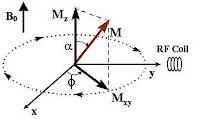
Thus, the already aligned nuclei (parallel or anti-parallel to the applied steady external magnetic field B0), has now been perturbed owing to the ‘knocking’ by the ‘magnetic component’ (B1) of the electromagnetic RF pulse. The nuclei gain energy and sway away from the perpendicular to the horizontal (90 degree) depending on how long the RF pulse is applied. So, now the nuclei behave like ‘punch-drunk’( like a person who’s been reeling due to a strong blow to the head!). magnetization vector resolved into its components horizontal Mxy and a vertical MzRemember, that this new angular momentum is also a vector quantity having magnitude and direction. It can be resolved in terms of a horizontal component (Mxy) and a vertical component Mz. Anyway, the proton does recover from this situation, after some time, once the external RF field has stopped. Typically, Mxy component decays faster than the recovery of Mz.

The excited proton recovers in two ways and both forms occur simultaneously: (1) The excited nuclei which now have been ‘forced’ to lie horizontally (90 degree), ‘re-align’ themselves back to their ‘original position’ as they were before the RF pulse (perpendicularly towards the field of externally applied field B0); and (2) the energized protons dissipate their energies to the surrounding nuclei (horizontally) at their level. The first example, obviously, is called the (spin-lattice, or longitudinal) relaxation; while the second one, transverse relaxation (T2). There is little energy loss due to RF emission.

T1 relaxation, also known as, longitudinal relaxation or spin-lattice relaxation can be best understood if you see the following Youtube video. [The spiral trajectory, in this case, reminds me of the laser experiment I did to satisfy my lesser friends. Analogically, the trajectory would be such, if the power supply were switched off.] In T1 relaxation, the proton loses energy to the surrounding lattice, by interacting with nuclei in the lattice which are in vibrational, translational and rotational motion. Clearly, the surrounding nuclei (lattice) having the same (or nearly same) Larmor frequency will efficiently absorb energy of the excited proton, resulting in a tiny rise of temperature.

T2 relaxation (transverse or spin-spin relaxation) on the other hand, does not involve exchange of energy with the lattice.
The magnetic moments of the protons merely changes phase. Here, the nuclei exchange “quantum states” (kind of, what Einstein called ‘spooky action at a distance’): an excited nucleus (proton) will transfer its energy and relax, while the neighboring nucleus in the lower energy state that absorbs it becomes excited. This loss of phase coherence of spins can be clearly seen in this beautiful video.

It can be understood easily that T1 and T2 values would depend on the surrounding molecular environment (tissues, for example). Hence, the values differ in different tissues. Again, since Mxy decays faster, as described, it may be understood why T1 is greater than T2 (usually, T1=5T2). Both T1 and T2 contribute toward contrast in tissues. T1 relaxation time is the time needed for 63% of protons to return to their previous equilibrium state. Likewise, T2 relaxation time is the time needed for 63% of protons to become dephased owing to their interaction with nearby protons. The contrast, naturally depends on the water content of the tissues. Grey matter has about 10% more water than white matter and this creates a contrast. We can also create contrast by varying TR and TE times.

TR (Repetition Time) refers to the time gap at which consecutive RF pulses are applied; while TE (Echo Time) refers to the time delay between the applied RF pulse and its reception (echo). T1 weighted images (T1W) are produced by keeping TR and TE relatively short, while T2 weighted images (T2W) are produced by keeping TR and TE relatively long. Water molecules being relatively light spins much faster than the Larmor frequency, making energy transfer rather tough (exchanging of packets of energy becomes more efficient as the relative angular velocity narrows). Consequently, water has a long T1 time. Proteins and nucleic acids being rather heavy, spin slowly. They also have problem with energy exchanging, and thus have a long T1. Cholesterol, a medium sized molecule, precesses near the Larmor frequency, efficiently absorbing the energy and giving a small T1 value.Thus (fat) liquid cholesterol in craniopharyngiomas, a benign tumor, appears bright on T1W images (T1 being small, the rate at which RF energy is released is fast. Hence, the signal intensity in NMR is high).

Subacute hemorrhage also has shorter T1, due to the presence of paramagnetic iron in methemoglobin present in the tissue, hence high signal intensity. Cerebrospinal fluid (CSF), edema (collection of fluid in tissue space or ECF) having more water content have both long T1 & T2 relaxation time. They give low signal intensity in T1 (dark) but higher signal intensity (bright) in T2W images. T2W images are superior to their T1 counterparts in case of infarction, edema, demyelination etc. Contrast agents like the heavy metal Gadolinium, a paramagnetic substance, has been used to reduce both T1 and T2 times by introducing inhomogeneity in the magnetic field. Gadolinium is complexed (chelated) with a substance called DTPA to prevent toxic build-up inside body tissues. This gives high signal in T1W but a low signal in T2W. It (the complex) does not cross the blood brain barrier (BBB); but disruption in the BBB or parts of the brain where it is deficient (circumventricular organs), take-up the substance and affects relaxation properties.

Below is an MRI showing changes in Subacute Sclerosing Panencephalitis, a complication of measles. T1 and T2 weighted MRI scansNote: Panels A and C are T1-weighted images; B and D are T2-weighted images. The hypointense (darker) signal on the T1-weighted image (arrow in A) and a hyperintense (bright) signal on the T2-weighted image (arrow in B) can be clearly seen.

Given all these, it can be said that relaxation parameters of nuclei have enabled us in visualizing biological tissues nonivasively, identifying chemicals spectroscopically and a lot more as we shall see later.

Last Modified: Aug 19, 2010

June 24, 2010

Understanding the Basic Principles of Nuclear Magnetic Resonance Imaging

ResearchBlogging.orgNuclear Magnetic Resonance Imaging (NMRI), better known as Magnetic Resonance Imaging (MRI) in medical parlance, is an invaluable tool in the study of the neurological system, soft tissue and musculo-skeletal system disorders. The word “Nuclear” was intentionally dropped later, as the procedure could then be wrongly interpreted by patients in relation to “ionizing radiation”, which certainly is not the case. However, the term Nuclear Magnetic Resonance (NMR) continues to be used in other (non-medical) fields of science, such as analytical chemistry, physics, biochemistry, petroleum industry, analysis of biological samples etc. In either case, the procedure and the basic principles remain the same. Paul Lauterbur was one of the pioneering inventors of this seemingly tough technological field.

Animation of a Helium atom
Matter is made up of atoms, which in turn, are composed of negatively charged electrons orbiting around the nucleus (look at the animation of a Helium atom on the left), consisting of positively charged protons and charge-less neutrons (with the exception of Hydrogen 1H nucleus, which contains a single proton and no neutron). These subatomic particles (electron, proton etc) somehow, can not be understood in terms of shape or color; instead they are denoted by their charge, mass or spin (angular momentum). An even number of them will cancel each other’s spin [just like two revolving spheres, in touch with each other would, in a ‘classical world’ (if one rotated clockwise, the other would rotate anticlockwise, canceling any resultant spin)].

Hence, a net resultant spin would result in the nucleus only if it contained an odd (unpaired) number of protons, an odd number of neutron or both. [The concept that certain nuclear species had angular momentum was first suggested by Wolfgang Pauli, while explaining the fine structures in the Atomic spectra. In the presence of an external magnetic field, the spectral lines got split, depending on the strength of the field (Zeeman Effect).]

Since nucleons bear a net charge (owing to the protons contained), the spinning nuclei will generate a magnetic field (since moving charges generate magnetic field). Each of these charged spinning ‘spheres’, hence, may be thought of as a tiny bar magnet having a magnetic dipole (that is a north-south orientation). [Electrons, similarly, have their own angular momentum though, responsible for molecular structure which nature uses, but they are not used by humans (Milestones in Spin podcast)] When we talk about “MRI” in humans, we mean proton nuclear magnetic resonance; i.e. NMR that detects the presence of hydrogen (proton) nuclei.

Our bodies have a plentiful of Hydrogen atoms: from the water within us, in cells and in extracellular fuid, (and to a lesser extent to the adipose tissue (fat)). These charge-carrying ‘unpaired’ protons (Hydrogen nuclei) rotate around their axes, but since all are spinning in a random fashion (as there’s no coordinator of any sorts); their net spin is zero, or in other words, their net magnetic moment is zero (as shown on the left).

Understanding spins aren't easy either. But, Prof. Stephen Hawking made it quite simpler for us using the real classical world analogy of ‘playing cards’ in his famous book A Brief History of Time (follow the link to learn more about ‘spin’). Having said that, the unpaired, positively charged protons having half integer (1/2) spins, behave like magnetic dipoles; it may now be understood easily that the spinning protons (nuclei) would align themselves to an externally applied magnetic field.

Precession of protons, animation

Thus, in a static magnetic field, the randomly oriented ‘tiny bar magnets’ align themselves up according to the applied magnetic field. These spinning protons (nuclei) also precess (make an angle) with the applied magnetic field (Bo), much like a spinning top does when its angular momentum diminishes. An animation of a proton precessing around a field is shown on the right.

[The magnet used for this purpose employs superconductivity. In a superconductive magnet, the electromagnet coils are immersed in liquid Helium at minus 269 degree Centigrade. At such a low temperature, the coils loose ‘resistance’ to the flow of electrons, resulting in a highly stable and a very strong magnet. (However, any minute vibration in the superconducting magnet can lead to runaway Eddy current leading to a phenomenon called 'quenching', that happened in the Large Hadron Collider at CERN, collapsing the whole setup.) Normally, 1.5 Tesla magnets are used, though nowadays 7 Tesla magnets have arrived. A 1 Tesla (1 Tesla=10,000 Gauss) magnet is 20,000 times stronger than the earth’s magnetic field)]. Also, note that we are considering magnetic moments along the axis of the external field only, as far as the sum-total alignment of individual magnetic torque contributing to a 'macroscopic' magnetization (M) is concerned. This is because the transverse components of the individual spins cancel out, as is seen in the 'cone' of the above picture. 600 persons of equal power, each pulling a rope either 30 degrees Northwest or 30 degree Northeast (in a 2 dimension), will certainly cancel out the 'east-west' vector, while the Northward vector will add-up. [It is this M that produces the induction current in the receiver coil].

The protons have two choices. Either they have to align parallel or anti-parallel to the applied magnetic field (known as spin-up and spin-down position respectively). In any case, the protons only ‘partially polarize’ since they tend to ‘make an angle with the applied static magnetic field. Spin down position is the higher energy state while spin-up state is the lower energy state of the spinning protons alignment of protons in a spinning nucleus in a static magnetic field(in the case of 23Na, there can be 4 spin-states instead of 2 as in 1H). (Obviously, a swimmer swimming upstream has more energy than his antiparallel counterpart.) The protons revolve (precess) around the direction of the magnetic field (Bo) at an angle, while at the same time they rotate around their own axis. Just as what happens in the solar system. [However, the upper (-1/2) and lower energy (+1/2) spin states are almost equally populated with only a very small excess in the lower energy state at room temperature. Since, there are so many of them that we finally make some headway].

Let me clarify a bit. You've seen a spinning-top rotating around its own axis. Due to Earth’s gravitational field, the top ‘maintains’ an angle (with the perpendicular/vertical), more visible when its angular momentum (speed) decreases, as it continues spinning. The top may be seen to revolve around “the perpendicular” at an angle (=‘precess’), (in addition to its rotation around its “own axis”) during its course of revolution. [Watch the Video "Introductory NMR & MRI Video 01 Precession and Resonance" to see what precession in NMR is]. This is what precession is about.

The frequency of precession is given by the Larmor relationship:
f=w/2*pi=yBo/2*pi (2*pi=360 degree)
w=angular freq. in radians per second; since there are 2*pi radians (360 degree) in a circle; we can find f, the frequency of rotation.
y is the magnetogyric (gyromagnetic) ratio, nuclear constant characteristic of every isotope. For 1H it is 42.5 MHz/T;
Bo=static magnetic field

The above equation is important, as we shall see later. Now let’s summarize what we learned so far.
Protons (nuclei) spin randomly in an atom. They tend to align with respect to an external magnetic field. These protons make an angle with the magnetic field as it goes about the magnetic field (while it also dutifully goes around itself), some parallel, and some antiparallel.

In MRI, our objective will be to disturb this alignment of protons with a dose of radio frequency pulse, in a similar way I discussed in my radio transmitter article but in a much, much bigger way. But since the ‘target’ (proton) is moving (precessing) around the field, we better ‘punched’ the target as if we were moving at the same angular velocity (so that the relative velocity was zero). Thus, when we apply the RF frequency pulses at the Larmor frequency, perpendicular to the magnetic field; the magnetic component (B1) of this electromagnetic wave temporarily knocks the protons out of alignment (see picture). If energy is absorbed by the nucleus, then the angle of precession will change. Assuming the field strength to be 1 Tesla, the protons are revolving 42.5 million times per second; it is at this frequency we give the pulse (i.e. at the Larmor frequency).

The protons are pushed out of alignment and as the pulse ends, they ‘relax’ (more on how they ‘relax’, later) back to their undisturbed ‘equilibrium’ position. This causes emission of an RF signal (the Echo) that can be picked up by the receiver coil (the same transmitter coil that produced it, in most cases); a damped oscillating wave generated, as the ‘disturbed’ magnetic moments coming back to realign with the magnetic field. Now, the problem begins. We have applied a uniform/homogeneous magnetic field (Bo). There are a lot of protons but we don’t know who’s who and residing where. That is why we also apply orthogonal magnetic field ‘gradients’ along the three (x, y, z) axes. [In a classroom, spray gradually ‘more’ yellow color in the front row and to the left than the back and to the right. In a similar way, spray blue color; hope your students don’t object. Now, every one of your students has a unique color: yellow, blue or green and with different hues]

Now that we get a decaying signal, which of high frequency; we mix it with a low frequency signal, in much the similar way as in heterodyning, to produce an ‘interferogram’. This interference map is digitized, which is called the Free Induction Decay (FID). Thus, we do find too many frequencies in ‘the low frequency map’ which occur in ‘almost’ the same time. So, what can we do?

Waka Waka! In this football World Cup 2010 at South Africa, audience seems to have a deafening organ, what they call ‘vuvuzela’. How are we going to analyze so many vuvuzelas when they are blowing at the ‘same time’? Just plot them in ‘frequency domain’ instead of ‘time domain’. Here’s Discrete Fourier Transform (DFT) which will do happily for you. [Simply put, it samples the different frequencies and plots them; not all vuvuzelas have the same frequency]

Now, that fuzzy picture of multiple frequencies has a 'spatial information' (owing to its orthogonal gradient magnetic field), contrast information (due to its ‘relaxation’ parameter), and foremost that it can be analyzed visually by humans, have enabled MRI to be a indispensable tool for the medical professional, as much as NMR has to the physicist or the discerning chemist. In MRI (NMR) it is not that important where or how energy is absorbed, but how quickly the excited protons revert back to its previous position is much more important, and hence the relevance of T1 and T2 relaxation times.

By the way, contrast depends on the t1 and t2 relaxation, the surrounding chemical environment affecting relaxation, and of course the water content of the tissues [gray matter contains 10-15% more water than white matter.]

Finally, the article wouldn't be resourceful enough if I do not post some MRI scans of the brain, this time, that of an epileptic patient (below).
(A sagittal section is obtained as the 'slice' takes a 'left to right' view (and vice versa); a coronal section means a 'front to back' view (or vice versa), and an axial slice means a virtual transverse section through the head.) Here's the picture of an actual MRI Machine below:

Naturally, the small tunnel may induce claustrophobia; the whirring acoustic noise from switched gradient coils may be troublesome to the patient; any implanted pacemaker may be subjected to interference from the electrical field resulting in dislodgement or malfunction (as in other ferromagnetic objects such as wrist watch, key rings etc.). Moreover, sudden movement by the patient may induce voltage in semicircular canal producing vertigo, a sensation of giddiness. Advances in MRI technology is happening fast. Claustrophobia may now be ameliorated with a wide bore MRI. A newly developed MRI scanner with Total Imaging Matrix (TIM) technology patients don't feel as claustrophobic, the imaging time is quick, quality of picture is better and even the acoustic noise is less (watch the YouTube video here). Whatever be the shortcomings of MRI, the benefits far outweigh the risks and it is here to stay and evolve.

References:
Magnetic Resonance Imaging: David D. Stark, William. G. Bradley, Jr.
NMR spectroscopy
Magnetic Resonance Imaging
MRI basics
Principles of NMR
William P. Dillon. Neuroimaging in Neurologic Disorders. In: Harrison's Principles of Internal Medicine, 17th Ed., Volume 2, McGraw Hill; 2008. p. 2491-2497.
Ian L. Pykett, Ph.D., Jeffrey H. Newhouse, M.D., Ferdinando S. Buonanno, M.D., Thomas J. Brady, M.D., Mark R. Goldman, M.D., J. Philip Kistler, M.D., & Gerald M. Pohost, M.D. (1982). Principles of Nuclear Magnetic
Resonance Imaging Radiology
P.S. We will discuss T1 and T2 relaxation, fMRI, tractography and NMR spectroscopy later.

Created: Jun 24, 2010; Last modified: Mar 10, 2014

March 24, 2008

The Medical Physics Of X-Ray

X ray of Roentgen's wife's handWilhelm Conrad Roentgen was passing a current through a 'partially' evacuated glass tube from an induction coil. Induction coils typically produce large make and particularly break currents due to collapsing magnetic field that produces electricity in the opposite direction. He noticed that a fluorescent screen glowed, despite the room being dark and the glass tube covered by black paper. He later noticed that this device produced some hitherto unknown 'rays' that could penetrate variety of materials. He could see through his own flesh, down to the bones, and medical application of X-ray was born. Later he captured this in a photographic plate. The photo on the left is that of Rontgen's wife's hand, still wearing the wedding ring, one of the earliest recorded X rays.

When there is a vast potential difference in a partially evacuated glass tube, physics of X rayelectrons rush towards the anode from cathode. As they impinge on the anode (anode being positively charged, attracts electrons), which is usually made of tungsten (Wolfram), they suddenly decelerate by colliding with the electrons of the anode material. Atoms are made up of protons and neutrons (which constitute the nucleus); and electrons, revolving around them in defined orbits. The electrons have clearly defined shells, spins, orbitals so that Pauli's exclusion principle is obeyed. Upon colliding, the electron knocks out an electron from the inner shell, which jumps to a higher energy level (outer shell). It finally comes down to its original place and emits electromagnetic radiation (photon) or X-Ray in the process. Thus X rays can be said to be produced mechanically (gamma rays are the result of nuclear decay or disintegration). The frequency of radiation is dictated by the equation, e=hv; where e is the energy of the quanta, h the Planck's constant and v the frequency of radiation.

X rays are electromagnetic radiation whose frequencies are higher than ultraviolet rays, but lower than gamma rays. They can be thought of as packets of energy or photons. X rays are ionising radiations. They ionize by either of the three ways: photoelectric effect, Compton effect, or pair production. In diagnostic applications (= at low energies, 30-100keV) photoelectric effects, the process just mentioned, predominate.
Photoelectric effects are proportional to the cube of the atomic number that is exposed (Z^ 3). This explains the mechanism why bones (containing calcium) contrast so well with soft tissues. At higher energies, as is employed in radiotherapy, Compton effect, whereby the incident electron transfers some of its kinetic energy (to impart) to the target electrons and the rest as a deflected, less energetic photon. At still higher energy (above1.02 MeV), the energetic electron will form 'matter-antimatter' pair in the exposed material. A positron and an electron will form, which will annihilate later (to form two photons which will fly almost 180 degrees apart, i.e. in opposite directions).

In addition to detection by photographic plates, fluoroscopy; solid state materials like lithium doped germanium or silicon can also detect x rays. Here, the photons cause the formation of electron hole pair, which can then be detected. X ray photons may be transformed into visible photons when they interact with alkali halides such as sodium iodide (NaI). This visible light may then be amplified by photomultiplier tubes. Strictly speaking, they are electron multipliers, because as x ray photons hit the halide surface, typically only a few atoms thick, electrons are emitted. These electrons are accelerated by subjecting them to a cascade of increasing positive voltages (dynodes) in a circular or venetian blind system. Photomultipliers require less x-ray exposure (due to this magnification) and are widely used in nuclear physics.

X rays have various uses in medicine, industry and science. Orthopedic situations such as fracture, joint displacement; cancer; lung parenchyma and associated illnesses like emphysema, pneumonia; calculi (stones: renal, gall stones etc); paranasal sinuses (PNS, in sinusitis) are among them. In physiology, biochemistry and many other fields, X-ray crystallography is a valuable armament in deciphering the structure of crystalline molecules. The photons diffract (scatter) as they travel through the crystal lattice, leaving its imprint in the process. One can decode the molecular structure by deciphering the 'scatterings'. Deducing the structure of DNA molecule is perhaps the best known example till date.

Natural structures can also emanate X rays. Some celestial bodies emit x rays. But don't ask me about X ray specs, I have no idea. Anyway, X-rays are not always invisible themselves. High energy x-rays, make the peculiar sensation of light produced within the eye itself, when someone looks directly into the beam.
References: Stephen M. Hahn, Eli Glatstein, "Environmental and Occupational Hazards", HARRISON'S PRINCIPLES OF INTERNAL MEDICINE, Vol.2 (15th edition) pp. 2586-2587.

ResearchBlogging.org
CrossRef DOI Query.
Last modified: never
Reference: hyper-links, unless specifically mentioned

May 06, 2007

Crazy Little Thing Called Life

sub marine lifeScientists have recently discovered a body of evidence regarding the existence of water on the red planet and has, for obvious reasons, gone gaga over it. They are conjuring up all the possibilities of existence of life on it.

Well, what are the basic ingredients of life? Are carbon, hydrogen, nitrogen and similar molecules extremely essential (mandatory) for life? What constitutes life and above all, how is life defined? If prions, (which are nothing but misfolded proteins, the cause of a myriad of illnesses like mad cow disease, kuru, scrapie and various other diseases) can be counted as living organisms, then perhaps any complex molecule or even radioactive elements like Polonium 210, which disintegrates on its own, to form many other elements, which in turn disintegrates (reminds me of Iron Maiden's 'Seventh son of a Seventh son' song), may be said to have life too, for they undergo an automatic activity. If you pore into the inside of an atom, you can see interactions in the nucleus: mesons; orbiting electrons around them, dutifully obeying Pauli's principle and many such activities that mimic life. Or are they living things themselves?

( En passant: Polonium 210 is found in tobacco in minute quantities; it was used to poison the famous Russian political dissident Alexander Litvinenko).

Life on earth perhaps needed the carbon skeleton, or it may even have been due to a chance occurrence. Microorganisms have been discovered in conditions in places, hitherto considered inviable for life (Bacillus stearothermophilus, B subtilis, Thermus aquaticus for example). The constituent of life could be molecules other than the conventional ones, even anti-matter! If we zoom in, we find tissues, cells, microtubules, mitrochondria, nucleus, the DNA and various other things which themselves are teeming with life. Zooming further still, we enter the constituent molecules to find the hadrons and the leptons, deeper still, quarks and gluons etc etc. They all seem full of life to me.

Look at the celestial objects. The sun is said to be a middle-aged star. So? Heavenly objects sometimes die a violent death: in supernova, or turn into neutron stars or black holes or some 'dwarf's. And the universe is said to have been born in the form of a major birth pang called the Big Bang. Are they living things then?

We know that there is a very small probability of finding two exactly similar humans, having all identical attributes. Likewise, no two electrons orbiting the nucleus can have the same quantum state, as per Pauli's exclusion principle (teleportation using entanglement/twiddling is an exception, for here two particles at a distance have the same properties) . This only shows that like unique human minds, the not-so-living things can also have their own uniqueness. Can they be said to have a life too, for they too move, have mass and energy. What about a cellphone or a computer or anything having AI(Artificial intelligence).

Let's be introspective. You are as much life as I am and we are made up of molecules arranged in a particular configuration: just matter; but where is the life? It may be here, in the entity called consciousness. Consciousness may be explained in terms of interactions among material elements. When we are dead, what exactly is missing? We may be brain dead, but the transplant surgeon may take out the kidney or cornea to transplant on others. Thus even after we die, we still continue to live in some of our tissues: they remain alive!

The question of life has to be addressed holistically, if we want to arrive at a sane and unanimous conclusion. Till then controversy will rage.