Showing posts with label neuroscience. Show all posts

September 07, 2010

The World of Tractography Where The White Matter Tracts Appear Colored

The Central Nervous System (CNS) communicates with the exterior (sensory e.g. gets visual, tactile information etc. on the one hand; and motor, performs limb movement, posture regulation etc. on the other) via the peripheral (somatic) nervous system. It also connects with the interior (our viscera or organs) via the Autonomic Nervous System. That is, it does its job in a bidirectional way: by the motor or the actuator arm, and sensory or the receptor arm. For such ‘actions’ to occur, cables of nerve fibers are laid within our body. Wouldn't it be nice if we could visualize these cables, their dispositional anatomy or any pathology that could afflict them?
a neuron showing an axon wrapped by myelin sheath
Schematic of a 'peripheral' myelinated axon 
The brain and spinal cord together constitute the CNS. We also know that there are about a hundred billion neurons in the CNS. Each neuron has a cell body (soma), an axon wrapped by myelin, and many dendrites. (See figure). It is the axon that carries the information in the form of action potential. These cables (bunch of axons, called tracts) are not laid haphazardly. Nature tries to conserve space, length, energy and so on and thus the axons form into tracts in a topologically efficient way. They run up and down (also front-back and sideways) the cord to the brain, the organ that we will now concentrate upon.

Your electrical wiring to your ceiling fan would include a switch and the fan itself. The wire (cable) from the switch would ascend vertically up the wall, make a 90 degree angle, and then reach the fan horizontally up in the ceiling. Likewise, in our brain, which is made up of two hemispheres, would connect. Three broad fiber types are seen: from one hemisphere to the other (commisural fibers), restricted to one hemisphere (front to back or antero-posteriorly are association fibers) and finally vertically (up down orientation go the projection fibers).

Exploring the tracts can now be done in live animals including humans. Improvement in MRI technology has enabled us to see the tracts (tractography). Improved gradient coils, faster processors and superior software have shortened the scanning time, thereby reducing 'blur' due to organ movement (e.g. diaphragm) and patient movement. This procedure called Echo Planar Imaging (EPI) has given birth to Functional Magnetic Resonance Imaging (fMRI), Diffusion Weighted Imaging (DWI), tractography and many other diagnostic and research procedures.

Consider the neuron shown in the above picture. Water molecules in the axon (yellow) are constantly in Brownian (random) motion due to thermal energy within. Hence they tend to diffuse constantly to come to equilibrium. In most of the cerebrospinal fluid spaces these microscopic motions are equal in all directions. This is called isotropic diffusion. But in myelinated neurons, as in the white matter fiber tracts, water motion is constrained due to the fatty nature of the myelin sheath (in blue) which hinders water flow across it. This anisotropic diffusion allows the flow of more water molecules along (parallel) the direction of the nerve fiber. The apparent diffusion coefficient (ADC) is thus more along the nerve fiber. Diffusion Weighted Imaging (DWI) can capture this microscopic water flow and delineate anatomically the orientation of nerve fiber tracts.

Pyogenic abscesses hinder diffusion by virtue of their increased viscosity, a rheological property. In the early stages of acute cerebral infarction there is reduced diffusion too, giving rise to high signal intensity. However, in most pathologies of the brain the ADC is increased. Diffusion Tensor Imaging (DTI), a diffusion MRI technology, tracks fiber orientation by assigning values in ellipsoid voxels (VOlume piXEL). Ellipsoid because unlike isotropic diffusion where molecules diffuse equally in all directions, the anisotropy in the white matter tracts does not permit them to move with equal ease in all directions, and hence the pattern is that of an ellipsoid and not spherical. By connecting the long axes of all the ellipsoids, the trajectory/orientation is deduced.

Colors are added to it with respect to the three principal axes (x, y and z)colorful world of tractography and the result is a stunning tractography! (see left)


The YouTube video below describes the colorful realms of tractography and how they are used.

When fMRI and DTI are combined together, a whole new world emerges. But, I prefer to keep it on hold till I discuss Functional Magnetic Resonance Imaging.

NB: The picture of the neuron/axon shown is not representative of CNS neurons. Oligodendrocytes that form myelin in the CNS does not encircle so many times as the Schwann cells in peripheral neurons do. It has been shown here only for the sake of clarity.

Last modified: Mar 10, 2014
Reference: Diffusion Tensor Tractography: Exploring the Cost-Benefit Ratio of Incorporating CSF Suppression into Fiber Tracing Algorithms

William P. Dillon. Neuroimaging in Neurologic Disorders. In: Harrison's Principles of Internal Medicine, 17th Ed., Volume 2, McGraw Hill; 2008. p. 2491-2497.

P. Mukherjee,, J.I. Berman,, S.W. Chung,, C.P. Hess, & R.G. Henry (2008). Diffusion Tensor MR Imaging and Fiber Tractography: Theoretic Underpinnings AM J Neuroradiol DOI: 10.3174/ajnr.A1051

November 20, 2008

Hearing Involves Sound Physics

Ear showing ossicles, round window and oval window
The way we hear sound is complex. The different attributes of sound (namely, intensity, frequency, the direction from which it is coming etc.) are faithfully perceived in the auditory cortex. The whole procedure may seem rather straightforward, but it is far more complicated than what looks so deceptively simple.

The sound waves (say from an orchestra) impinge on our eardrums. Sound waves are mechanical waves consisting of condensation and rarefaction, things we learned in our school days. These waves then vibrate our eardrums (Tympanic Membrane; TM). The TM is critically damped, meaning any vibration that is set in will stop almost instantaneously. The vibrating TM then transfers its mechanical energy to the oval window, in the membranous labyrinth of the cochlea, via an ossicular chain consisting of three (3) very small bones.

Basilar membrane, organ of Corti and the scala vestibuli,media and tympani, phalangeal cellsThe membranous labyrinth consists of three adjoining tubes coiled side by side (as shown in the figure). If we were to make a section through it, we would find 3 separate compartments within it: Scala vestibuli, Scala media and Scala tympani. Scala vestibuli is connected to Scala tympani at the apex of the cochlea, a place called helicotrema. While Scala tympani contains a fluid called endolymph (a fluid rich in K+ or potassium ions); the other 2 tubes contain perilymph (a fluid very similar to plasma, rich in Na+ and low in K+). The sheer asymmetry in K+ distribution among the two adjacent fluids (endolymph and perilymph) generate an endocochlear potential of about +80mV, endolymph positive when the perilymph is considered as 0 (zero) volt or ground.

As the oval window vibrates, the fluid in Scala vestibuli (perilymph) also vibrates. Sound was traveling in air before it struck the eardrum, but here, we see that they are now propagating in a fluid medium, which has far more inertia than air. The possible impedance mismatch that would happen is compensated by the eardrum itself and the ossicular chain. The mechanical advantage of the lever system of the ossicular chain, together with the ratio of surface areas of TM and the 'oval window', amplifies the force of sound waves about 22 times, so that the total force at the oval window is 22 times than what the TM experienced originally.

Now, vibrations have set up in the Scala vestibuli form the oval window. These vibrations find their way to the Scala media, as the two tubes are separated by only a very thin membrane (Reissner’s membrane). Hence, fluid in the Scala media (endolymph) vibrates whenever the oval window is vibrating. This Youtube video beautifully illustrates it.


The vibrating endolymph sets up a wavy motion in the basilar membrane (BM). The ‘real analysis’ of sound waves starts here! The BM performs real time spectral analysis of sounds it is presented with (analysis of frequencies below 200Hz is skipped though). We normally hear in the frequency range of 20Hz to 20 kHz.

The hair cells in the organ of Corti, our hearing apparatus, are arranged in such a manner along the BM that those near the base of the cochlea will respond to high frequencies; while as we go up to the apex of the cochlea, the BM reacts best at low frequencies. In other words, each part of the BM has its own unique maxima, the frequency at which the BM responds most well. Below 200Hz, there is no such place encoding.

traveling wave in the basilar membrane
Our ears follow another principle. The ‘traveling wave’ spreads quicker near the base of the cochlea and its speed diminishes fast as it goes up. This ensures that a longer stretch is available for the higher frequencies; else the higher frequency part would have been bunched together, creating a loss in the HF range. This non linearity in traveling wave propagation is thus needed.

Generator Potential
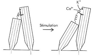
Tip links of hair cells of the earThe genesis of generator potentials in the hair cells is also interesting. Imagine that a group of persons of varying height are standing on a carpet. A thread is attached from the top button (of his shirt) of the smaller person to the top button of his taller counterpart. If the carpet is now tilted by pulling it up from the short person’s end, the thread will now be stretched snapping the taller person’s button. The hair cells also have threads (Tip Links) extending from shorter to their taller cousins. A traveling wave will cause pulling of tip links, resulting in the opening of a mechanically sensitive cation channel. Since potassium is the predominant cation in the endolymph, K+ will then enter the taller hair cells. Look at the electrical gradient around the hair cell; it is -140mV with respect to the endolymph (it is about negative 60 mV wrt the perilymph). So, cations, specially K+ rushes in  creating depolarization. A cascade of events like opening of voltage-gated Ca++ ion channels at the base, consequent fusion and exocytosis of vesicles discharging neurotransmitters, probably glutamate to the afferent cochlear nerve endings surrounding the hair cells, creating an action potential in the nerve.

Frequency Discrimination
When we listen to music, our ears pick up the frequencies in a number of ways. Firstly, the place (maxima) on the BM where maximum excitation takes place is actually a function of frequency. As a matter of fact, there is a frequency map along the BM. Secondly, at frequencies below 3 kHz, the nerves fire in synchrony with the incident sound waves. This is called the ‘volley principle’. The ‘phase locking’ of the two frequencies that occurs below 3 kHz, is highly analogous to the ‘phase locked loops’ in electronic circuits (NE565). Thus ‘volley principle’ allow us to discriminate frequencies. Actually, volley effect is more important in ‘loudness’ assessment. ‘Pitch’ (the subjective/psychological dimension related to frequency) are also moderated by factors such as loudness and the duration of sound. At low frequency (below 500 Hz) pitch seems lower and at higher frequencies (above 4 kHz) pitch seems higher, as the loudness increase, when the frequency is kept constant. Again, when the duration of sound increase from 0.01 second to 0.1 second, the pitch will rise too, for a particular frequency. Sound of less than 0.01 sec duration does not evoke appreciation of any pitch by us.

Loudness Discrimination
Loudness is the perceived intensity of sound, a subjective psychological dimension. The interpreted sound sensation is proportional to the cube root of the actual sound intensity. As such, the ear works at the top of its limit, at a point analogous to Hopf bifurcation, beyond which instability in oscillations occur.
As the sounds become louder, the amplitude of BM movement is more, resulting in more excitation of hair cells. Secondly, with greater loudness, the hair cells around the ‘maxima’ fire too. This causes spatial summation. Thirdly, the outer hair cells are stimulated at loud sounds. The brain will automatically infer loudness levels when cells corresponding to the outer hair cells fire.

Locating Sounds
Then there is location of the direction of sound. We can locate whether the drums are on the left and the lead guitar is to the right. This is achieved by calculating which ear gets the sound first (time lag) and/ or which ear gets it louder (intensity). The time lag method works below 3 kHz; while intensity method works at higher frequencies.
Front/ back discrimination is done by the pinna (auricle) of our ears due to their particular shape.

Auditory nerve
It is interesting that each auditory nerve fiber has its own characteristic frequency, the frequency at which it responds most well. However, it is true only at low intensity. At higher intensities, this specificity is lost and they then respond to a wider spectrum of frequencies. The auditory nerve produces a flurry of action potentials, the frequency of which depends on the intensity of the sound stimuli, it is exposed to. This is very much similar to ‘voltage to frequency converter’ ICs (LM331 is one such VFC IC), where a change in voltage at the input of VFC will cause a change in frequency at the output.

The auditory nerve then goes to the: cochlear nucleus in the medulla to Superior olivary nucleus to Inferior colliculus (via lateral lemniscus) to Medial Geniculate Body (in the Thalamus) to end in Auditory cortex. Some fibers cross to the other side early in their course while others cross at other levels. Fibers from primary auditory area sends association fibers to different parts of the brain for language processing and other tasks.
The nerve synapses with higher order neurons in the above places, which then relay to the nerves upstream. Everywhere in its course, including the nuclei, there are clearcut ‘tonotopic maps’, representing definitive frequency layouts. There are also extensive crossing of nerve fibers to the opposite side. Before the fibers reach the auditory cortex, they connect to many reflex pathways vital to life. For example, they send branches to the reticular activating system which keeps us awake during the noisy hours.

The auditory cortex (Brodmann’s area 41) is the portion of the cerebral cortex in the superior temporal gyrus. Its anterior part is mainly concerned with low frequency and the posterior part tackles the higher frequencies. Thus area 41 also has its own tonotopic map. Secondary auditory cortex (auditory association area) is vital for the interpretation of sounds. A person, in whom Wernicke’s area (part of auditory association area) is damaged, will hear normally but will fail to understand its meaning, leading to aphasia.

The sheer complexity of the auditory circuitry is really mind-boggling: the logarithms (we hear in a log scale, not a linear one), cube functions, phase locking are only some of them. The range of sound intensity (from whisper to the roar of a jet plane) we hear is about 1 trillion fold; but surprisingly, the auditory nerve fibers have a much less dynamic range. Yet we hear the full range. It’s really amazing.

ResearchBlogging.org
P. Martin (2001). Compressive nonlinearity in the hair bundle's active response to mechanical stimulation Proceedings of the National Academy of Sciences, 98 (25), 14386-14391 DOI: 10.1073/pnas.251530498

Last modified: Mar 10, 2014
Reference: Textbook of Medical Physiology, 17e, Guyton and Hall

November 22, 2007

Stem Cell Therapy for Paraplegics May be Near

picture of SupermanOur beloved superhero, the Superman, played by Christopher Reeve is no more. He fell from a horse and damaged his spinal cord in the process. As a result he got paralyzed and was restricted to the wheelchair. He could have recuperated if only the cut ends of the spinal cord were repaired. He was a staunch advocate of stem cell research.

It is now known that the spinal cord harbors stem cells, cells capable of differentiating into nerve cells (essentially all types of cells). So where have the stem cells gone? In adults, a molecule called netrin1, directs the stem cells away from an injury. Modifying the behavior of netrin1 may help in the recruitment of stem cells and their much needed transformation into neural elements. Simple grafting is impracticable, as cross connection may spell doom. Auriculotemporal (Frey syndrome) is one example where faulty neural connection gives rise to unpleasant symptoms. Moreover, the nerves and their synapses are so spatially oriented that correct axial positioning is next to impossible. For example, neurexins are molecules which are found in the synaptic terminals, which plays a pivotal role in the alignment of a synapse. They form connexions with neurexin receptors situated in the post synaptic neurons and thus guides the presynaptic terminal. The neural networking is indescribably complex.

Finding a solution through netrin1 may be of immense benefit not only in spinal cord injury but also in diseases like cerebrovascular accident (CVA/ Stroke).

Again, the brain-computer interface robots being developed by researchers, based on 'thought controlled devices' are becoming a reality. These humanoids may one day enable paraplegics to be able to move freely in their wheel chairs, command computers or TV stations remotely and in other day to day activities.

November 15, 2007

Brain Sees What The Eyes Can Not Behold:The Strange Facade of Optical Illusion

card showing misspelled textIf you look carefully at the image on the left and read the text aloud, chances are that you will read this SMS type text just as gracefully as you would read ordinary texts. Despite the several spelling mistakes that the eye detects, the brain comes in to fill in the void, by its own damage control machinery. Optical slips are also managed in more or less the same way when we see TV or watch a movie, which are typically delivered in frames per second.

There is no denying the fact that the physiology of visual sensation itself is interesting. Hyperpolarization of photoreceptors plays important role in the propagation (generation) of nerve signals. It is in stark contrast to what happens normally; depolarization of cells as a means of signal generation and propagation. Moreover, the image retina gets, as a result of light passing through the lens, is an inverted real image. But we see them normally, not upside down, as a result of some manipulations by the brain.

autostereogramThe picture on the left is an example of autostereogram. This is a FANTASTIC picture! Just save the picture in your computer, then open it using Windows Picture and Fax Viewer. Look at it but don't focus on the plane of the picture! Easier said than done. Now try to adjust your focus back and forth; a picture of a 3 dimensional ball will begin to emerge. Concentrate on it. When you can behold the image relatively easily, for a longer period, zoom in on the image, using the scroll button of your mouse. A brilliantly colored spirally twisted cone will advance towards you, perpendicular to the plane of your computer monitor's screen. Its really tough, initially, but exercise your patience, you will get the experience of your lifetime. You can see some more of these stereograms here.

Many other interesting optical illusions are there, and some even take into account, the 4th dimension: the images move. You can get some of them here. However, sometimes the eyes see right but the brain goes bonkers. In the extreme example of visual illusion, one interprets a coiled rope for a snake. This may have been the result of the survival advantage it offered, which we acquired in the course of evolution. So, trick the eye and fool the brain.

October 23, 2007

Stroop Task and The Anterior Cingulate

Stroop TaskTry to read (name) the colors in which the words are written and not the words themselves. See how you stumble and fumble. Beg your pardon, everyone stumbles at this seemingly minuscule task. This test is aimed to make you fumble. This testing procedure, called the Stroop Task, actually tests the conflict between the lower and higher areas in our brain. Here, lower and higher mean hierarchically (functionally, evolution-wise) lower and higher respectively and NOT anatomically.

In my previous article, 'Right is Might', I discussed about how the left and right cortex did their chores in their own unique ways. Now, the part of the brain that is involved in this conflict, lies midway between the two cortices, called the anterior cingulate cortex. This little area of the brain connects the two cortices so that information can flow bidirectionally, between the two hemispheres. We are pretty adept in understanding the SMS (short messaging service) text jargons from mobile phones, even though the words are misspelled; can see continuous motion pictures, even though they are actually shown at 48 frames per second. This highlights our instinctive ability to decipher them easily. On the other hand, naming colors (not merely seeing them) is more resource intensive and is taken care of by higher brain areas. Thus in a task like this, where two threads of activity occur simultaneously (reading the texts and naming the ink in which they are written) an interference or conflict is produced. And hence the slowing down. It can be compared to bottlenecking due to multitasking in computer systems. In fact, it is through the anterior cingulate, system buses from both upper and lower functional areas, in addition to, right and left cortex (anatomically) pass.

The anterior cingulate not only decides the bone of contention but also determines error related negativity (ERN). That is it somewhat functions like a "differential operational amplifier" of electronics, in which the output signal is the difference between input signals . By doing this, it may be able to instruct the motor system which task is to be given precedence. It also acts as seat of emotion. For example, in (lactating) women her baby's cry will elicit strong emotions on her. This has been studied by fMRI (functional Magnetic Resonance Imaging). It may also explain the fact that women are more socially sensitive.

August 19, 2007

Recent Advances in Memory Research

RAM chipsThough how memory is formed, stored or retrieved has not been elucidated yet, things are beginning to unfold as new advances begin to break the frontiers. It was long thought that long term memories (memories such as the persons own name), which remain steadfastly seated in the brain, were etched permanently in the brain. But, recent advances in research has proved to the contrary. Long term memories are not like something as a computer's ROM (Read Only Memory, which once stored can not be erased and it does not need any maintenance current once the data had been entered; EPROM and EEPROM are exceptions), but it resembles a computer's RAM (Random Access Memory, which needs to be continuously fed with electricity in order to retain its memory).

In a classic experiment by Prof. Yadin Dudai, Head of the Department of Neurobiology at Weizmann Institute of Science, Israel, and colleagues had trained rats to learn to avoid certain tastes. This is called conditioned avoidance, a form of operant conditioning (learning that is influenced by environment). This is done by giving an unpleasant stimulus such as electric shock or injecting substances that cause discomfort, along with some foods. Thus the animals 'remember' to avoid these foods which are associated with bad accompaniments. Prof. Yadin Dudai had injected a chemical, an inhibitor of a protein called PKM zeta, into the rats' taste cortex. A single administration was found to erase the long term memories of those rats completely. The rats forgot which tastes they were to avoid.

PKM zeta (or Protein Kinase M zeta) is an isoform of protein kinase C, an enzyme present within the cell membrane. This molecule (PKM zeta) is necessary for an already learned and deep rooted response (long term memory) to remain long term. The constant ferrying activity of PKM zeta in the synapse (junction between two neurones) is needed for the memory to be retained, reminding us of the constant supply of electricity needed in the RAM of computers to retain its memory. Thus the long founded idea that long term memories were etched permanently into the brain is now being challenged and a new dynamically modeled approach such as this is emerging.

A process called long term potentiation (LTP) occurs in the brain which help us to learn. Repeated high frequency stimulation of a synapse strengthens it by LTP. It should be noted in this context that nicotine helps in memory formation since it stimulates the inhibitory inputs to layer V pyramidal cells. These cells exert a negative influence on LTP based learning. Hence suppression of these cells by nicotine helps in memory formation.

Another very useful discovery is the development of a new microscopic technique that has allowed us to 'see' memory traces in the brain. Using restorative deconvolution microscopy, scientists at the university of California at Irvine, has shown how the synapse had expanded after LTP. If you think of the synapse as the space between two balloons, you will realize that by pressing these balloons towards each other, the balloon ends will flatten, leading to a broader synapse with a larger contact area. Just as in Ohmic resistance of electricity, here also the apparent resistance will diminish, leading to a faster and better processing and storing.

Such long giant leaps are needed to achieve better nootropics (memory enhancers), treat and cure Alzheimer's disease and related conditions.

July 27, 2007

Meditation and Physiology

person meditatingMeditation is the process of concentrating upon one's own self, fathoming deep down into the labyrinth of one's own consciousness. Scientists wanted to see whether meditation had any demonstrable effect on the physiology of the performer. They took eight long term Buddhist practitioners (monks), who used to practice regularly, for this purpose. They also took ten healthy volunteers, who did not practice it, in the study (as a control group, for comparison and for judging the statistical relevance of data.)

They recorded the electroencephalographic (EEG) activity of both the groups and analyzed them. They found higher (statistically significant) gamma band activity in the monks, while they were in the act. Their relative baseline gamma band activity (25-42 Hz) over low frequency activity (4-13Hz), was also better. Both relative gamma power and absolute gamma power were greater in them, than the controls. Not only that, long distance gamma synchrony in them was far greater.

Since it is known that gamma synchrony plays an important part in the formation of consciousness [1, 2], it can be only inferred that they stay more alert and attentive than the control population. Also, long distance gamma synchrony, being more well developed in them, it is only natural that the integration intensive functions like memory, intelligence, and other cognitive faculties are better developed. Its no mambo jumbo, the research has been thoroughly studied with established scientific methods and substantiated statistically. No wonder, that transcendental meditation helps patients of AIDS or hypertension.

Meditation can be beneficial to our mental, spiritual and overall physical help. It is a great de-stressor.

Ref:
Long-term meditators self-induce high-amplitude gamma synchrony during mental practice
Antoine Lutz, Lawrence L. Greischar, Nancy B. Rawlings, Matthieu Ricard, and Richard J. Davidson
doi: 10.1073/pnas.0407401101

[1]. http://citeseerx.ist.psu.edu/viewdoc/download?doi=10.1.1.119.2469&rep=rep1&type=pdf

[2]. http://link.springer.com/article/10.1007/s10867-009-9148-x

June 16, 2007

Consciousness: An Inside Story

person meditatingIt is a daunting task to explain what is consciousness, for there is no consensus in this regard. Though many different definitions exist; it can simply be defined as the feeling of ones own lively presence with in oneself. How this feeling is brought about in the living beings, including humans, is not precisely known and many a hypotheses have been propounded. The most appealing as well as the most tenable among these is that it is due to the processes and interactions that occur in the hydrophobic domains of dendritic proteins in the neurons of the brain. Proteins are chains of amino acids (like beads in a string) which have different shapes depending on what the constituent amino acids are. Amino acids containing hydrophobic side chains such as valine, isoleucine etc. and amino acids containing aromatic rings such as phenylalanine, tyrosine are (by definition) very much insoluble in water. Thus dendritic proteins which bathe in the extra cellular fluid consisting of water fold themselves in such a way so as to make an infolding, an inward pocket where these particular (hydrophobic) amino acids are located. It is with in this microcosm of pockets strange things happen.

As we all know, that in the nano world, weak forces such as van der Waals become increasingly relevant where as those of gravity begins to take a backseat. In these pockets an interaction occurs between these adjacent non polar (hydrophobic) molecules. Just like the south pole of a bar magnet would induce the formation of a north pole in a ferromagnetic substance there is the creation of opposite charges on the nearby molecule by the action of 'transient dipoles' (the hydrophobic molecules behave as transient dipoles in which charges oscillate/interchange between the poles. The charges occur as a result of imbalances in the electron clouds of the molecules). This attractive force that exists between transient dipoles is also known as London force, a kind of van der Waals force.

The oscillations that occur in the dendritic proteins are synchronized in a global scale with in the brain. As a result 'binding' occurs, which means that there is consolidation in a global scale within the brain about all the attributes of a thing. For example, when someone gets hurt he feels pain by the help of his thalamus and locates the painful stimulus by the help of his cerebral cortex. Binding helps us to feel both simultaneously, as shown in the example. It is necessary as the brain processes tasks parallely. It is much akin to the clocking in a computer to a fixed frequency.

Exactly how this clocking/synchronization is done is not known for certain. This synchronization is also known as the gamma synchronization as it is reflected as gamma waves in the EEG (electroencephalogram), having a frequency in the range of 30-70 Hz. This synchrony can occur as a result of electromagnetic coupling between regions within the brain and this would explain why it is done almost instantaneously. That the brain radiates electromagnetic waves is well known.
Another possibility is quantum coupling by the processes of entanglement. According to quantum mechanics it is possible for a particle to learn about another one, in no time, without ever having to go to that particle physically. This 'instantaneous/in no time' phrase is important as we do not want any phase lag to creep in so that a decoherence state does not occur. In the leaves of plants too, the electrons sparked off by photons/sunlight go hopping (by mind reading!) to the place where actual photosynthesis is taking place by previously reading the quantum state of other electrons by entanglement.

The association of hydrophobic pockets assumed with the formation of consciousness can be inferred from the fact that swamping these charges by external electric fields as in electro anesthesia, would result in 'unbinding' and anesthesia. Again, chemicals such as anesthetics which dissolves the hydrophobic pockets cause anesthesia by disrupting the London forces attempts to strengthen our notion.

References: The Brain From Top to Bottom
Quantum Consciousness & Orch-OR

May 14, 2007

The Cerebellum-Part2

start menu of Windows XPQuite a few of us are acquainted with the fact that in Windows XP, the start menu items that display, when we click the start button, organize themselves continuously (dynamically), as we use them. The programs we use most frequently, are placed on the top; while the least used items settle down at the bottom. A similar hierarchy is followed by the brain too. The tasks we have inherited from our ancestors, phyllogenetically, are kept at the bottom of our memory/program cache while more recent skills are placed at the top. As we go on acquiring new knowledge, they first locate in the cortical areas;- once the task have been mastered, they settle down deeper, into the cerebellum and so on. It seems we have our own stack registers. For example, much of our basic and posture maintenance algorithms are kept in lower brain areas such as the spinal cord, medulla, the midbrain rather than the cerebral cortex. They are old programs, evolution wise, and hence kept in the lower shelves. In early neonates, and in diseases of neuronal pathways e.g. in upper motor neuron lesion, midbrain lesions, spinal cord lesions the functional architecture/hierarchy is lost, as the higher centers are not yet or no longer active.That is, now, the sophisticated programs of the cerebral cortex are no longer working, the phylogenetically/ evolutionarily old programs resurface. Babinski reflex in UMN lesions, Moro's reflex are two examples.

In addition to the above mentioned address lines, there is this real time integration and differentiation that goes on constantly within the brain. The much awed calculus is their regular cup of tea!

To be contd.

May 13, 2007

Cerebellum: Electronically Speaking

an EPROM chipSometimes journeying into physiology textbooks can be a deja vu in electronics. The other day I was studying the way cerebellum learns from its past 'karma' and memorizes the best setting for an action. I was surprised to find that its learning resembled a lot with the op-amp (operational amplifier) ic's (LM741, for example) and its memorizing resembled to that of the EPROMs (erasable programmable read only memory, a picture of which is shown here on the left).

The cerebellum has one of the largest nerve cells of the body, the Purkinje cells, in addition to other types of cells. Purkinje cells have numerous tree-like branching dendritic processes. They receive two types of electrical connections; from the mossy fibers (about 250000 to 1 million fibers for each Purkinje cell) and climbing fibers (ONLY one for each cell).

The cerebellum fine tunes the movement that accompanies a certain task in the following way. The brain (lateral portions of the cerebellum and the basal ganglia) 'plans the action' even before we start an action. After the task has been executed, the cerebellum calculates the 'error', the difference between the planned trajectory and the achieved output in much the same way a does.
In the figure on the left, the op-amp is configured as a negative feedback, through resistance R2 which feeds a portion of output voltage back into its inverting input (since input is fed into its - terminal) amplifier. This way, the op-amp can have both a fraction of the output and input at the same pin, thereby the gain and other parameter remain 'stable'.


A still better analogy phase locked loop operation is that of the phase locked loop, the output phase of which 'locks' to the input frequency (diagram on the left). Likewise, the cerebellum too matches its expected action with that of the resulting action. The cerebellum is 'happy' with the performance when the error is minimum. At this moment of bliss, the climbing fibers fire for a long duration and with a characteristic waveform: a spike followed by a long trail. Now the information is written permanently.

Doesn't our experience of programming a 2716 (EPROM) tell a similar story; a peak programming voltage and a normal working voltage? When we wanted to program an EPROM, we needed to give it a high programming voltage in the beginning, followed by a steady 5 voltage thereafter till the EPROM 'learned'.

It doesn't end here. In the cerebral cortex, memory is consolidated in sleep, when it receives a spike voltage: the k-complex in the NREM stage of sleep. It seems that we have a micro, or may be a pico-controller in our brain.

To be contd.