 The heart is vital to our bodies, since it pumps blood into various parts of our bodies, that provide us oxygen, nutrients and takes away the metabolites for disposal. For this, the heart has to contract in order to generate enough force for the ventricles (atria too) to have a standard ejection fraction. Not only does it contract, it also generates the impulses necessary for its contraction, a property called automaticity, by which it generates its own rhythm.
The cells in our bodies are bathed in a sea of fluid. This fluid, called the extracellular fluid or ECF, as it is located outside the cells. The ECF is rich in sodium ions (Na+), while their concentration inside the cells are much less. Conversely, the concentration of potassium ions (K+) is more inside than outside. It is due to this difference in concentration of ions on either side of the cell membrane, a transmembrane voltage is produced. These ions being polar in nature and water soluble, can not penetrate the lipid cell membrane. But they can gain entry, through specialized pores called ion channels.
The sodium ion channels, channels through which sodium ions pass, has 2 gates: an activation gate and an inactivation gate. What controls these gates is not known for sure, but it could either be an energy barrier controlling its entry or a flexible peptide chain-like stuff that alters the conformation of the gates to restrict the ion's access. Extracellular calcium ions also restrict sodium ions to go through and thus has a stabilizing effect. These gates are called voltage controlled gates, since the opening or closure of the gates are controlled by transmembrane voltage. These gates are inactivated at voltages above -55mV (i.e. say -40 mV) and no sodium ion can pass. Below -70 mV , the gates are open, allowing free flow of Na+ along the electrochemical gradient, from the exterior to the interior of the cell.
There are areas of heart other than the sinus node which have intrinsic automaticity i.e. they are capable of generating their own rhythms, such as the Purkinje fibers. But the SA Node (sinoatrial node) is the normal pace-maker, since it fires at the highest rate. In the nodal tissue, where the cell voltage hovers around -60 to -55 mV (millivolt), the sodium ion channels are inactivated at this voltage. Since, the resting membrane potential (RMP) of the pacemaker lies in a region where the sodium channels are inactivated, the firing of the SA Node depends on other ions, particularly calcium ions (Ca++). These nodal pacemaker cells (possibly P cells, containing little organelles) are inherently 'leaky' to calcium ions.
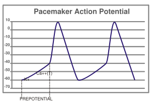
 So, Ca++ enters these cells making the cells' interiors less negative, (Ca++ is a cation bearing 2 positive charges. A cation is a positively charged ion, so called because it is attracted towards the cathode). Once the cell gains enough positive charge, to become a little more positive, about -40 mV, as shown in the figure (prepotential), there is a sudden spurt of calcium influx (impulse). T type (transient) calcium channels are responsible for the prepotential while L (long lasting) type calcium channels are responsible for the impulse. In addition to the influx of calcium ions from the ECF, calcium is also liberated locally from the sarcoplasmic reticulum of the nodal cells, known as the 'calcium spark'. So, the cell is now fully depolarized, as shown. But soon calcium channels close and influx stops. Potassium channels open. Since the concentration of K+ is more inside, K+ leaves the cell making the interior more negative. The efflux of K+ and stopping of further influx of Ca++ repolarizes the cell, making it ready for another cycle. Ca++ leaks again and the cycle repeats.
The colored animation on the top left shows the spread of cardiac impulse, from the SA Node. Click on the animation if it doesn't animate on its own.
Last modified: Mar20 2009
Reference: Basic and Clinical Pharmacology, Bertram G Katzung, 9th ed, page 220
No comments:
Post a Comment